
|
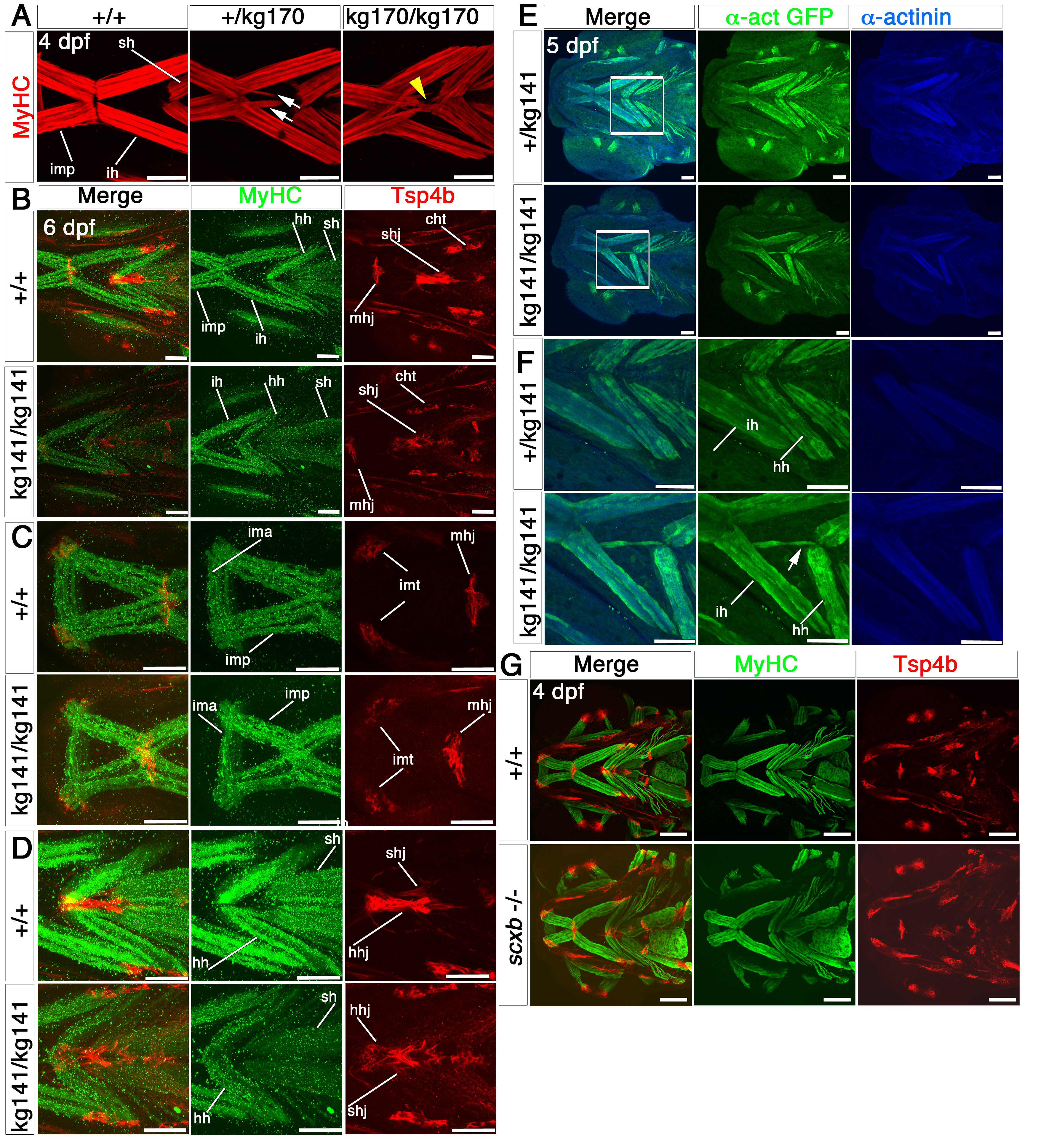
Fig. S3
Cranial tendons, ligaments and muscles of scxakg141 mutants are abnormal and disorganized. Confocal stacks of cranial muscles using immunofluorescence for MyHC (A4.1025, red in A), Tsp4b (red) and MyHC (MF20, green) in B-D, GFP (green) and alpha-actinin (blue) in E,F and Tsp4b (red) and MyHC (A4.1025, green) in G. All in ventral view, anterior to left. A. Embryos from a scxakg170/+ incross at 4 dpf; scxa-/- mutants had misaligned fibres and tri- and four-way abnormal junctions (yellow arrowhead). Some scxa+/- embryos had some milder defects (white arrows). B-D. 6 dpf embryos from a scxakg141/+ incross; the matrix protein, Tsp4b was downregulated in mutants, and some tendons such as the mandibulohyoid junction and intermandibular tendon (C), the sternohyideus and hyohyoideus tendons (D) were misshapen and showed decreased matrix condensation. Muscle fibre defects contained fibres connecting to wrong muscles and disorganized junctions. E,F. 5 dpf embryos from a scxakg141/+;Tg(actc1b:egfp)zf13 incross. F shows magnified boxed region in E. Mutant embryos had fibres from the interhyoideus muscle growing in the wrong direction towards the hyohyoideus junction (white arrow). G. Both tendons and muscles of 4 dpf embryos from a scxb+/- incross looked normal. mhj, mandibulohyoid junction, ima, intermandibularis anterior, imp, intermandibularis posterior, imt, intermandibular tendon, sht, sternohyoides tendon, hhj, hyohyoideus junction, ih, interhyoideus, hh, hyohyal, sh, sternohyoides. All scales 100μm except A, 50μm.