
|
Fig. S4
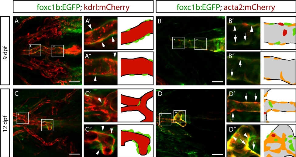
foxc1b:EGFP is expressed in vascular smooth muscle cells on the ventral aorta at larval stages. Ventral images of zebrafish embryos at 9 and 12 dpf. A) At 9 dpf, foxc1b:EGFP positive cells (arrowheads) are located adjacent to the kdrl:mCherry expressing endothelial cells of the ventral aorta (n = 4 embryos). B) At 9 dpf, foxc1b:EGFP and acta2:mCherry are co-expressed in mural cells (arrows) with the exception of a few single-positive cells (arrowheads; n = 4 embryos). C) At 12 dpf, foxc1b:EGFP positive cells continue to associate with endothelial cells of the ventral aorta (n = 6 embryos). D) At 12 dpf, most mural cells co-express foxc1b:EGFP and acta2:mCherry, although there are some single positive cells (n = 6 embryos). Grey colour in model indicates endothelial patterns. Scale bars represent 50 µm.
Reprinted from Developmental Biology, 453(1), Whitesell, T.R., Chrystal, P.W., Ryu, J.R., Munsie, N., Grosse, A., French, C.R., Workentine, M.L., Li, R., Zhu, L.J., Waskiewicz, A., Lehmann, O.J., Lawson, N.D., Childs, S.J., foxc1 is required for embryonic head vascular smooth muscle differentiation in zebrafish, 34-47, Copyright (2019) with permission from Elsevier. Full text @ Dev. Biol.