
Image
|
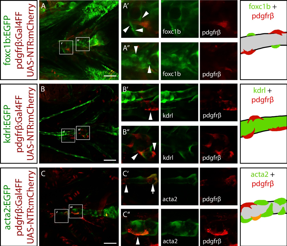
Figure Caption
Fig. S3
foxc1b:EGFP positive cells do not co-express a pericyte marker. A) foxc1b:EGFP is not co-expressed with pdgfrβ:mCherry in the ventral head. B) pdgfrβ:mCherry is associated with the endothelium (kdrl:EGFP) in the ventral head. C) pdgfrβ:mCherry and acta2:EGFP are partially co-expressed in ventral head mural cells. Schematics depict mural cell and endothelial marker expression that match the transgenes. Grey indicates presumptive endothelial patterns. Scale bars represent 50 µm.
Figure Data
Acknowledgments
This image is the copyrighted work of the attributed author or publisher, and
ZFIN has permission only to display this image to its users.
Additional permissions should be obtained from the applicable author or publisher of the image.
Reprinted from Developmental Biology, 453(1), Whitesell, T.R., Chrystal, P.W., Ryu, J.R., Munsie, N., Grosse, A., French, C.R., Workentine, M.L., Li, R., Zhu, L.J., Waskiewicz, A., Lehmann, O.J., Lawson, N.D., Childs, S.J., foxc1 is required for embryonic head vascular smooth muscle differentiation in zebrafish, 34-47, Copyright (2019) with permission from Elsevier. Full text @ Dev. Biol.