
|
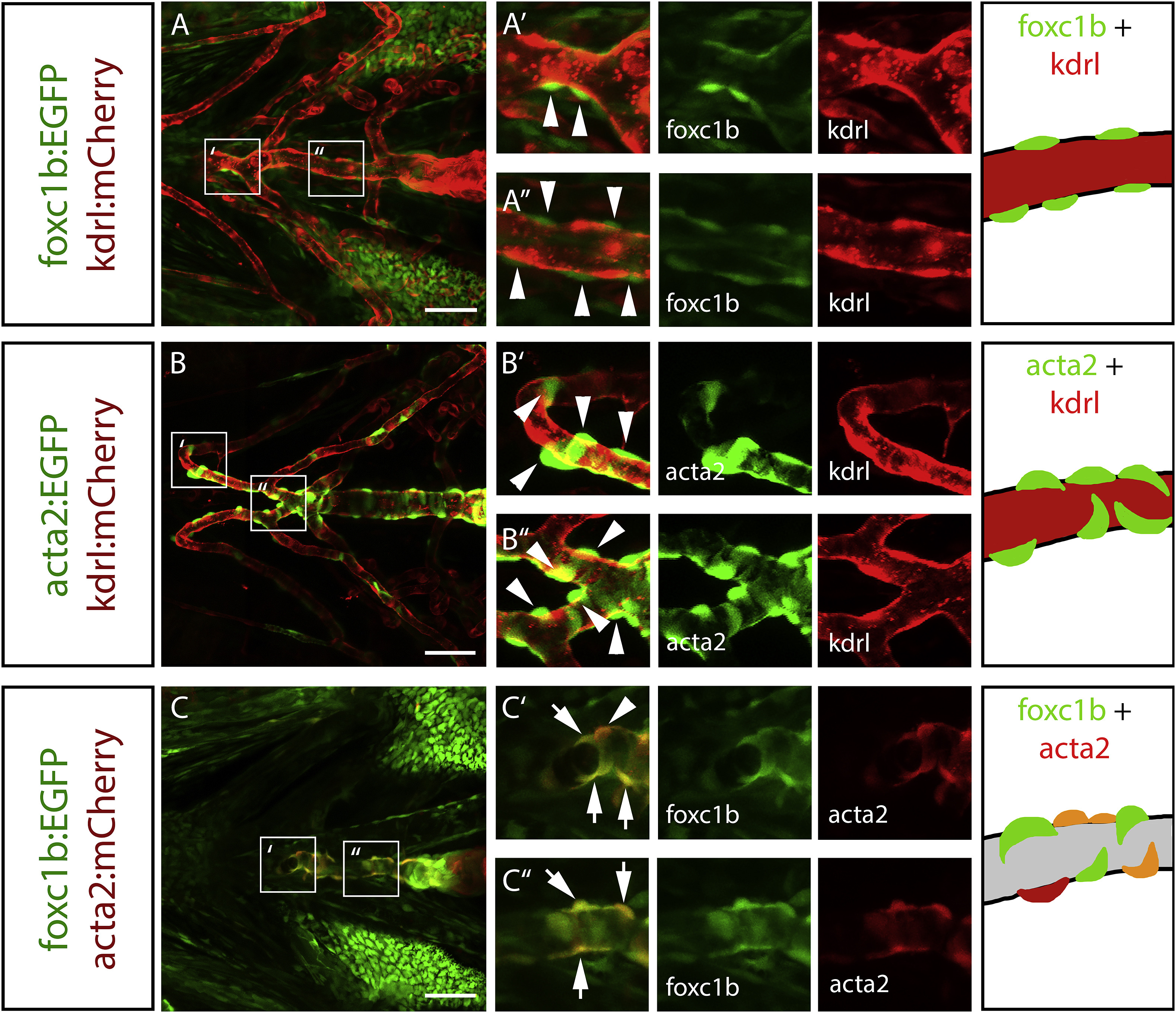
Fig. 3
foxc1b expressing perivascular cells in the embryonic ventral head co-express acta2. A) In ventral head vessels at 4 dpf, foxc1b:EGFP expressing cells are associated with, and surround, the endothelium (kdrl:mCherry) along the ventral aorta. B) Similarly, acta2:EGFP positive smooth muscle cells surround the kdrl:mCherrypositive endothelium along the ventral aorta and aortic arch arteries. C) foxc1b:EGFP and acta2:mCherry are co-expressed along the ventral aorta. Schematics depict mural cell and endothelial marker expression matching the transgenes. Grey indicates presumptive endothelial patterns. Arrowheads represent marker-expressing mural cells. Arrows show co-expression of two smooth muscle cell markers. Scale bars represent 50 μm.
Reprinted from Developmental Biology, 453(1), Whitesell, T.R., Chrystal, P.W., Ryu, J.R., Munsie, N., Grosse, A., French, C.R., Workentine, M.L., Li, R., Zhu, L.J., Waskiewicz, A., Lehmann, O.J., Lawson, N.D., Childs, S.J., foxc1 is required for embryonic head vascular smooth muscle differentiation in zebrafish, 34-47, Copyright (2019) with permission from Elsevier. Full text @ Dev. Biol.