
Image
|
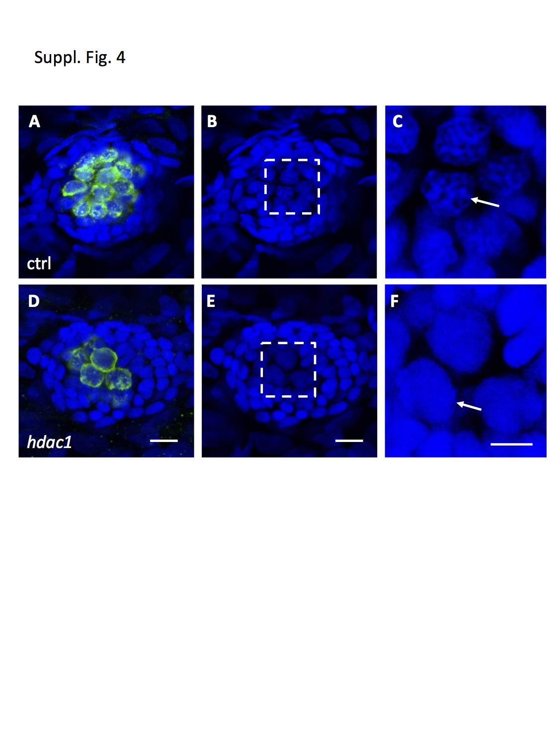
Figure Caption
Fig. S4
hdac1 mutation causes abnormal hair cell morphology.
(A-F) Confocal imaging of immunofluorescently-stained neuromasts in the control (A-C) and hdac1 mutant (D-F) embryos. Hair cells were stained in green with hair cell-specific antibodies to myo6 and myo7, neuromast nuclear DNA were stained in blue using DAPI. C and F are the magnified images of the areas boxed in B and E, respectively. White arrows in C and F point to the morphology of hair cell nuclear DNA, which is more condensed in the control embryo (C) than in the mutant embryo (F). Scale bar, 10 μm in A, B, D and E; 5 μm in C and F.
Figure Data
Acknowledgments
This image is the copyrighted work of the attributed author or publisher, and
ZFIN has permission only to display this image to its users.
Additional permissions should be obtained from the applicable author or publisher of the image.
Full text @ NPJ Regen Med