
|
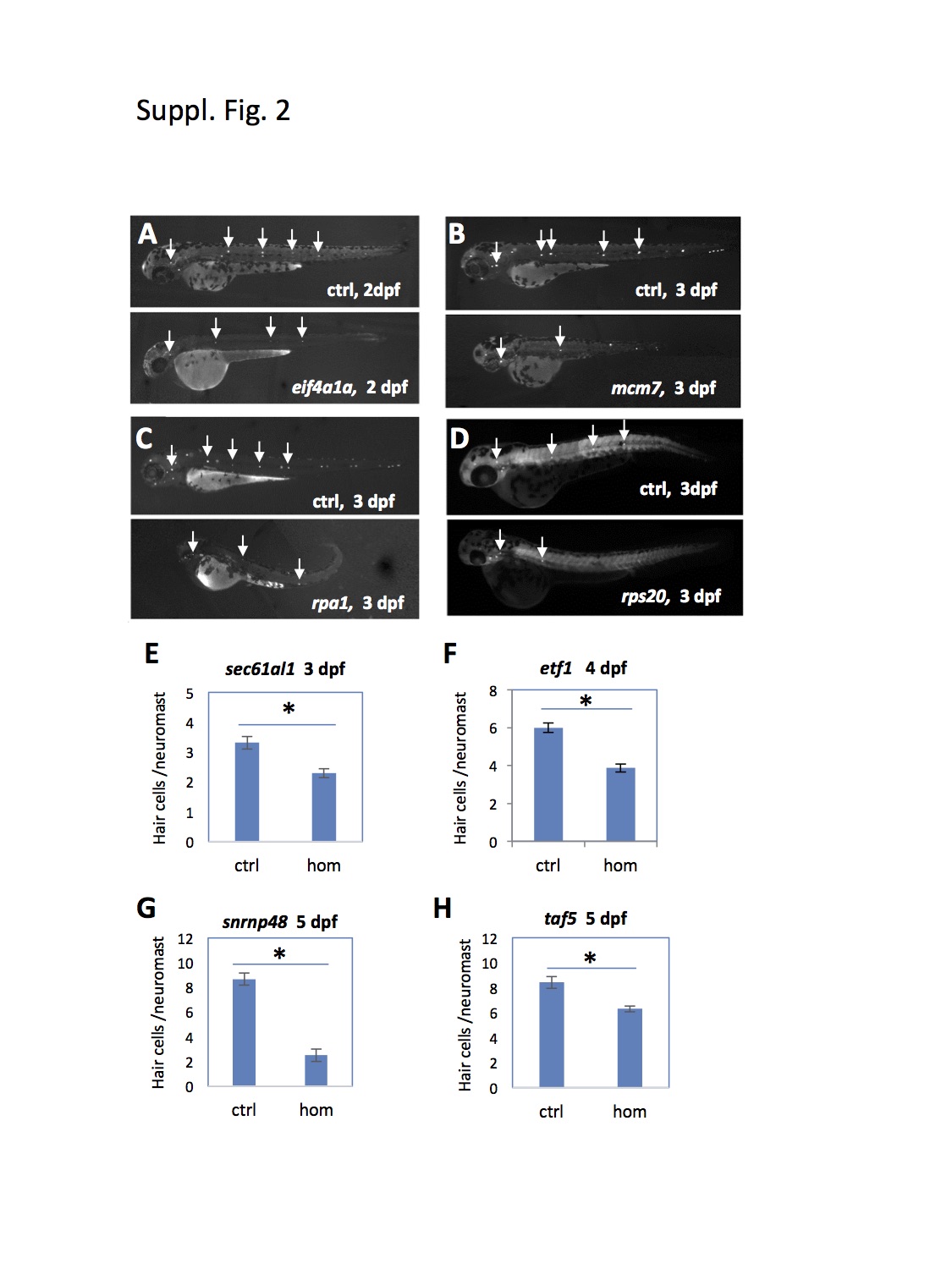
Fig. S2
Mutations tested that affected hair cell development.
(A-D) Developmental timing of neuromast hair cell formation was assayed by Yopro-1 staining (A-C) or hair cell-specific antibody staining (D). Arrows point to hair cell-positive neuromasts. Mutant embryos had smaller staining area for each neuromast and fewer neuromasts, indicating an impairment in hair cell development. (E-H) Quantification of neuromast hair cell development by counting Yopro-1 positive hair cells. Compared to the control (Ctrl) siblings, homozygous (hom) mutants of sec61a1show a significant reduction in the average number of hair cells per neuromast.