
|
Fig. 5
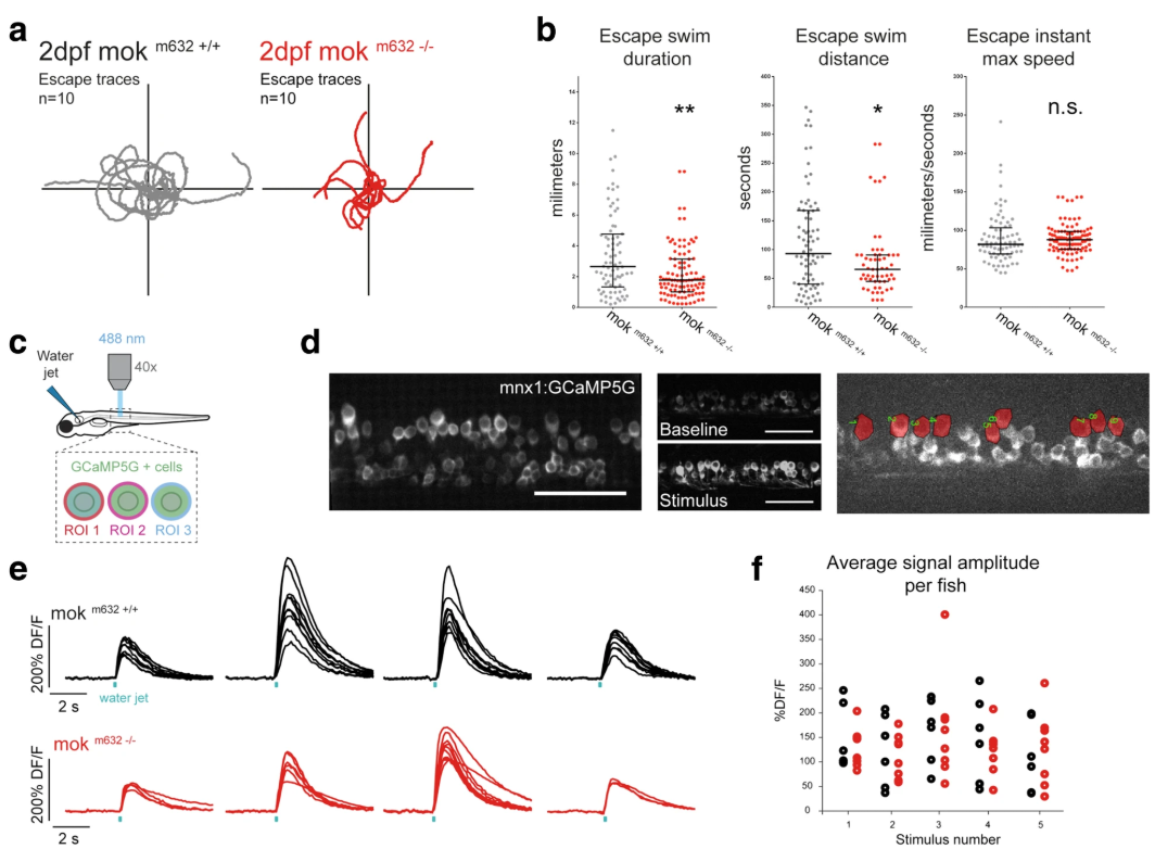
NMJ dysfunction leads to behavioral deficits. a NMJ functional defects lead to impaired locomotor behavior in 2dpf embryos as determined by touch-evoked escape response assay. Escape traces extracted from video tracking of escape swimming episodes following the presentation of a stimulus for 10 embryos per genotype shown here as an example. bQuantification of escapes reveal that Dynactin1a depletion leads to impaired locomotion determined by reduced escape duration and distance, but without altering maximum instant speed. c Calcium imaging of fictive escape responses in motor neurons expressing GCaMP5 was performed in the spinal cord upon presentation of a water jet stimulus. d GCaMP5 expression was confined to motor neurons and analysis of calcium signals was performed on dorsally-located primary motor neurons (region of interest in red). e Example of calcium signals obtained from primary motor neurons including CaP motor neurons in mok m632−/−larvae (red) and their wild-type siblings (black) at 4dpf; one trace per cell, four fictive escape responses are represented to show response variability. f Maximum DF/F amplitude signal in dorsal motor neurons averaged per fish and plotted according to the stimulation number, showing proper recruitment of spinal cord motor neurons despite reduced levels of Dynactin1a. Data shown as b) median +/− interquartile range (b: n = 76,101; f: n embryos/n cells = 6/63, 8/44) Scale bar 100 μm