
|
Fig. S2
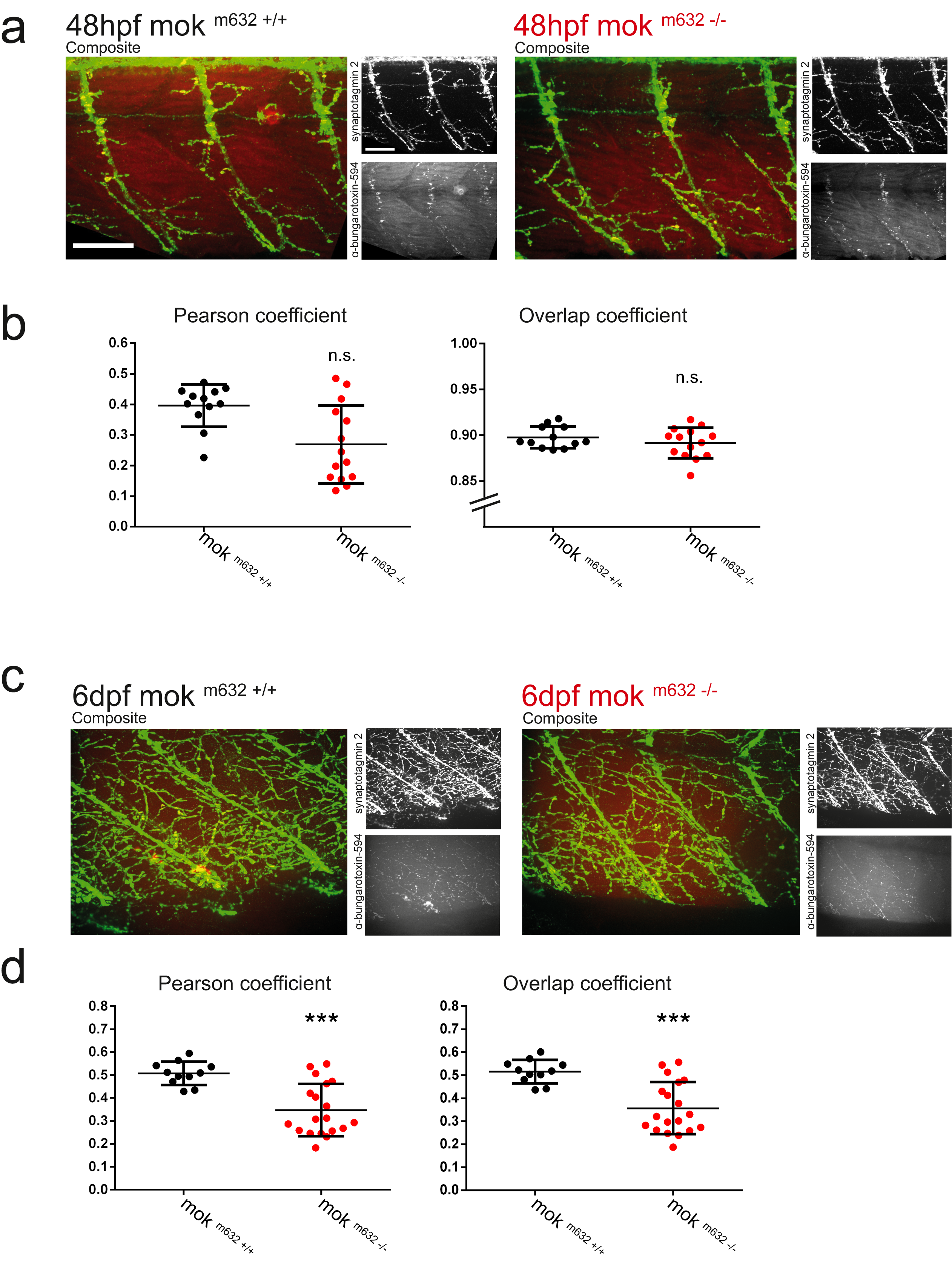
mok m632−/− embryo and larvae NMJ structural integrity. a) Double immunohistochemistry reveals the integrity of the NMJ at 2dpf by coverage and colocalization of presynaptic structures (anti-synaptotagmin2, in green) and postsynaptic Ach receptors (α-bungarotoxin, in red). b) Quantification of the colocalization shows normal NMJ structure of the ventral root at 2dpf by both Pearson’s coefficient and the overlap coefficient. c) NMJ structure is also affected at 6dpf, with d) reduced coverage in pre- and postsynaptic components, as well as reduced colocalization. All data presented as average +/− SD; (b: n embryos = 12, 14; d: n larvae = 11,19) (TIF 32575 kb)