
Image
|
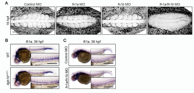
Figure Caption
Fig. S6
Itgb1b and Fn Are Dispensable for EC Specification, Related to Figure 5 and 6. (A) Confocal stack fluorescence images of 16 hpf wild type embryos injected with control MO, fn1a MO, fn1b MO, and both fn1a and fn1b, and immunostained with anti-Fn antibody. Dorsal views with anterior to the left. (B, C) Expression patterns of fli1a in 36 hpf sibling (WT) and itgb1bmi371 embryos (B) and in 36 hpf embryos injected with control MO and fn1a/fn1b MO (C). Dorsal views with anterior to the left. The boxed areas are enlarged in the insets. Scale bars: 100 μm (A); 200 μm (B, C)
Acknowledgments
This image is the copyrighted work of the attributed author or publisher, and
ZFIN has permission only to display this image to its users.
Additional permissions should be obtained from the applicable author or publisher of the image.
Reprinted from Developmental Cell, 49(5), Rho, S.S., Kobayashi, I., Oguri-Nakamura, E., Ando, K., Fujiwara, M., Kamimura, N., Hirata, H., Iida, A., Iwai, Y., Mochizuki, N., Fukuhara, S., Rap1b Promotes Notch-Signal-Mediated Hematopoietic Stem Cell Development by Enhancing Integrin-Mediated Cell Adhesion, 681-696.e6, Copyright (2019) with permission from Elsevier. Full text @ Dev. Cell