
|
Fig. S3
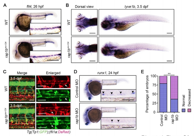
Rap1b Is Required for HE Specification but Dispensable for Specification of Venous and Lymphatic ECs, Related to Figure2. (A) Expression patterns of the venous marker flt4 in 26 hpf wild type and rap1bncv124 embryos. (B) Expression patterns of the lympathic EC marker lyve1b in 3.5 dpf wild type and rap1bncv124 embryos. Dorsal (left) and lateral (right) views with anterior to the left. (C) Confocal stack fluorescence images of trunk axial vasculature in 3.5 dpf sibling (WT; upper) and rap1bncv124 (lower) larvae in the background of Tg(Tp1:GFP);(fli1a:DsRed). Lateral views with anterior to the left. Merged images of Tp1:GFP (green) and fli1a:DsRed (red). Enlarged images of fli1a:DsRed and Tp1:GFP corresponding to the boxed areas are shown at the right. Arrows indicate thoracic duct. (D) Expression patterns of HE marker runx1 in 24 hpf embryos injected with control MO (upper) and rap1b MO (lower). Arrowheads indicate runx1-positive hemogenic ECs. (E) Percentage of embryos showing normal (3 or more, blue) and decreased (less than 3, pink) number of runx1-positive hemogenic ECs in the AGM regions as observed in D. Control MO, n=35; rap1b MO, n=51. **, p < 0.01. In (A), (C) and (D), the boxed areas are enlarged in the insets. Scale bars: 200 μm (A, B, D); 50 μm (C)
Reprinted from Developmental Cell, 49(5), Rho, S.S., Kobayashi, I., Oguri-Nakamura, E., Ando, K., Fujiwara, M., Kamimura, N., Hirata, H., Iida, A., Iwai, Y., Mochizuki, N., Fukuhara, S., Rap1b Promotes Notch-Signal-Mediated Hematopoietic Stem Cell Development by Enhancing Integrin-Mediated Cell Adhesion, 681-696.e6, Copyright (2019) with permission from Elsevier. Full text @ Dev. Cell