
|
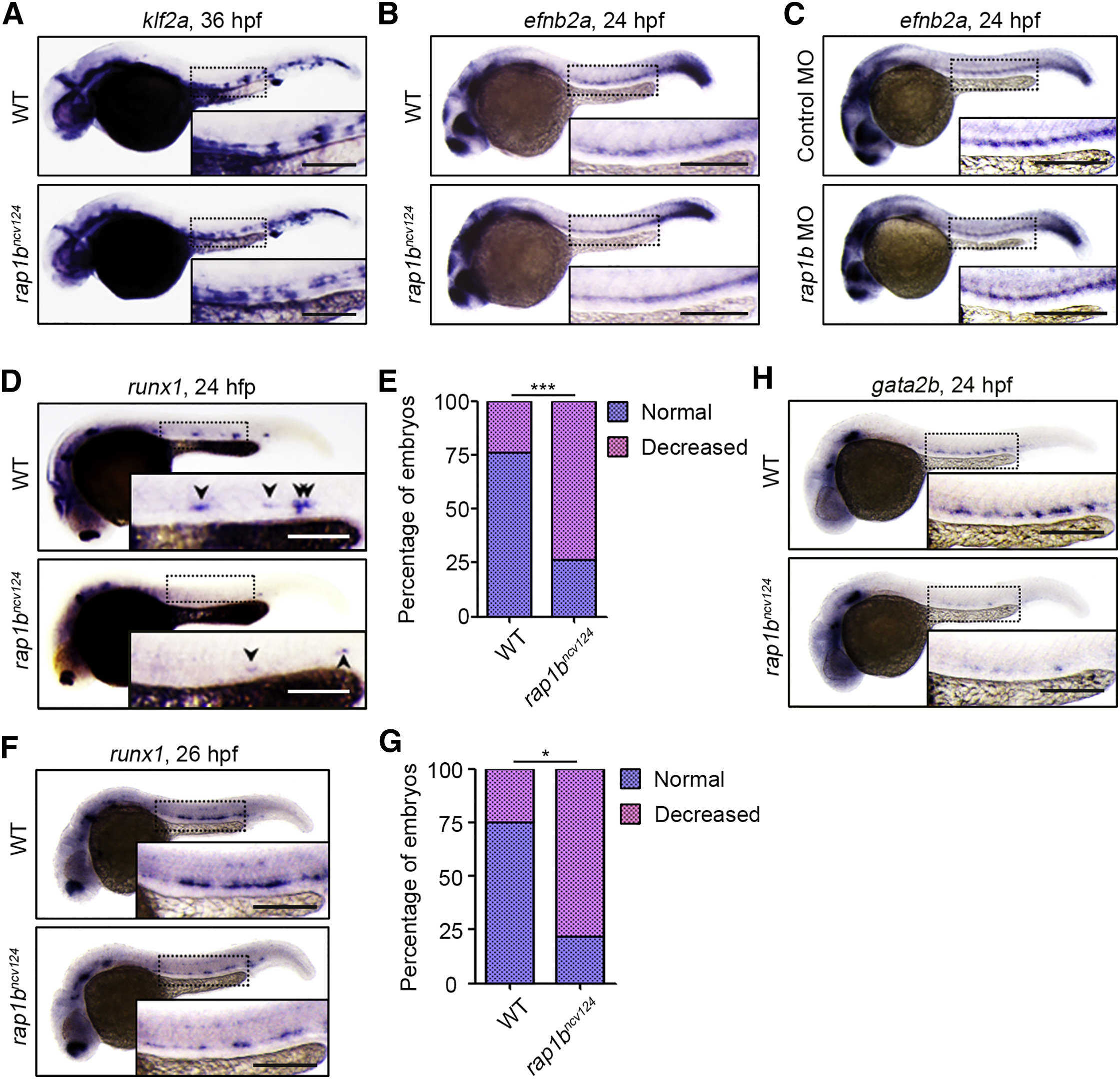
Fig. 2
Rap1b Is Involved in HE Specification
(A) Expression patterns of blood-flow-responsive gene klf2a in 36 hpf sibling (WT) and rap1bncv124 embryos.
(B and C) Expression patterns of arterial marker efnb2a in 24 hpf sibling (WT) and rap1bncv124 embryos (B) and in 24 hpf embryos injected with control MO or rap1b MO (C).
(D) Expression patterns of HE marker runx1 in 24 hpf sibling (WT) and rap1bncv124 embryos. Arrowheads indicate runx1-positive hemogenic ECs.
(E) Percentage of embryos showing normal (3 or more, blue) and decreased (less than 3, pink) numbers of runx1-positive hemogenic ECs in the AGM regions, as observed in (D). WT, n = 42; rap1bncv124, n = 43. ∗∗∗p < 0.001.
(F) Expression patterns of runx1 in 26 hpf sibling (WT) and rap1bncv124embryos.
(G) Percentages of embryos showing normal (blue) and decreased (pink) expression of runx1 in the AGM regions, as observed in (F). WT, n = 12; rap1bncv124, n = 10. ∗p < 0.001.
(H) Expression patterns of another HE marker, gata2b, in 24 hpf sibling (WT) and rap1bncv124 embryos.
In (A)–(D), (F), and (H), boxed areas are enlarged in the insets. Scale bars: 200 μm (A–D, F, and H)
Reprinted from Developmental Cell, 49(5), Rho, S.S., Kobayashi, I., Oguri-Nakamura, E., Ando, K., Fujiwara, M., Kamimura, N., Hirata, H., Iida, A., Iwai, Y., Mochizuki, N., Fukuhara, S., Rap1b Promotes Notch-Signal-Mediated Hematopoietic Stem Cell Development by Enhancing Integrin-Mediated Cell Adhesion, 681-696.e6, Copyright (2019) with permission from Elsevier. Full text @ Dev. Cell