
|
Fig. 7
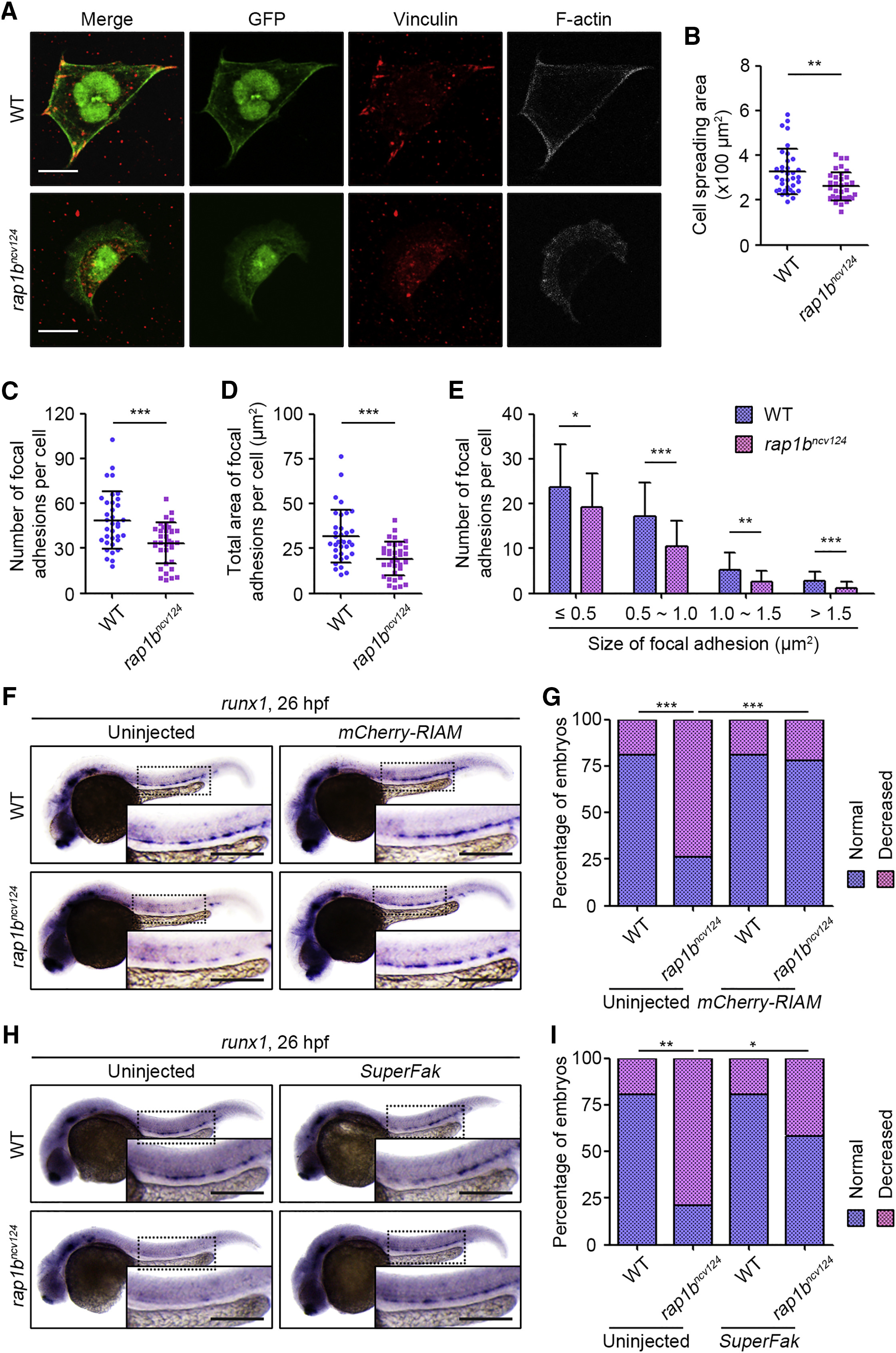
Rap1b Regulates HE Specification through Promotion of Itgb1b-Mediated Adhesion of PLPM Cells
(A) Cells dissociated from 17 hpf WT (upper) and rap1bncv124 (lower) Tg(fli1a:GFP) embryos. Merged images of GFP (green) and vinculin (red), GFP (green), vinculin (red), and F-actin (gray) images are shown as indicated.
(B) Spreading areas of GFP-positive cells derived from WT and rap1bncv124 embryos, as observed in (A).
(C) Number of vinculin-marked focal adhesions per cell, as observed in (A).
(D) Total area of vinculin-marked focal adhesions per cell, as observed in (A).
(E) Number of vinculin-marked focal adhesions, as observed in (A). Data are shown as mean ± SD.
In (B)–(D), each dot represents an individual cell. Error bars indicate means ± SD. In (B)–(E), WT, n = 35; rap1bncv124, n = 35. ∗p < 0.05; ∗∗p < 0.01; ∗∗∗p < 0.001.
(F) Expression patterns of runx1 in 26 hpf sibling (WT, left) and rap1bncv124 (right) embryos injected without (left) or with mCherry-RIAMmRNA (right).
(G) Percentages of embryos showing normal (blue) and decreased (pink) expressions of runx1 in the AGM regions, as observed in (F). Uninjected WT, n = 17; Uninjected rap1bncv124, n = 16; mCherry-RIAM mRNA-injected WT, n = 20; mCherry-RIAM mRNA-injected rap1bncv124, n = 14. ∗∗∗p < 0.001.
(H) Expression patterns of runx1 in 26 hpf sibling (WT, left) and rap1bncv124 (right) embryos injected without (left) or with SuperFak mRNA (right).
(I) Percentages of embryos showing normal (blue) and decreased (pink) expressions of runx1 in the AGM regions, as observed in (H). Uninjected WT, n = 15; Uninjected rap1bncv124, n = 14; SuperFak mRNA-injected WT, n = 15; SuperFak mRNA-injected rap1bncv124, n = 16. ∗p < 0.05; ∗∗p < 0.01.
In (F) and (H), boxed areas are enlarged in the insets. Scale bars: 10 μm (A); 200 μm (F and H).
Reprinted from Developmental Cell, 49(5), Rho, S.S., Kobayashi, I., Oguri-Nakamura, E., Ando, K., Fujiwara, M., Kamimura, N., Hirata, H., Iida, A., Iwai, Y., Mochizuki, N., Fukuhara, S., Rap1b Promotes Notch-Signal-Mediated Hematopoietic Stem Cell Development by Enhancing Integrin-Mediated Cell Adhesion, 681-696.e6, Copyright (2019) with permission from Elsevier. Full text @ Dev. Cell