
|
Fig. 2
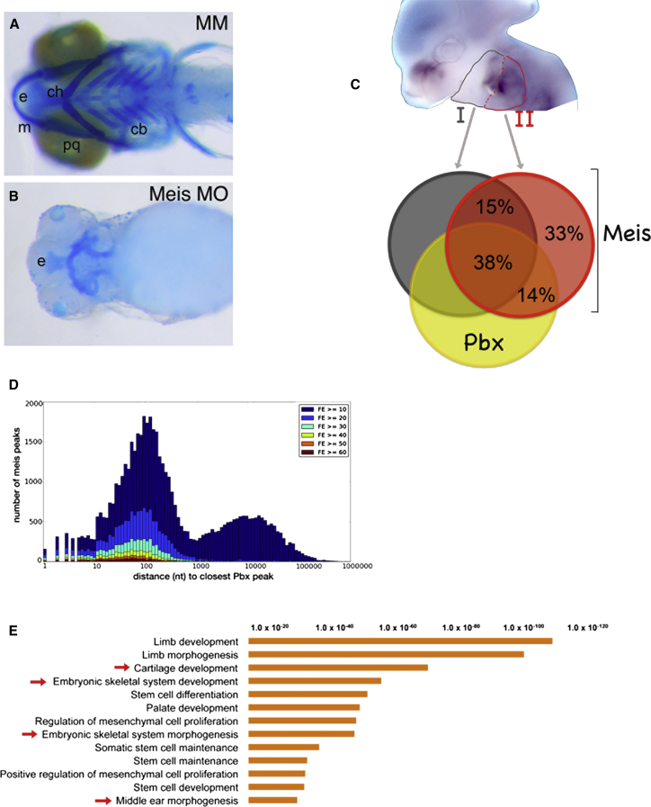
Meis TFs Are Required to Form the Branchial Arch-Derived Skeleton
(A and B) Ventral view of zebrafish larval (6 days postfertilization) control (mismatched morpholino, MM) (A) and Meis-morpholino-injected embryos (B) head skeleton. The IIBA-derived skeleton (ceratohyal) is labeled by ch.
(C) Craniofacial region of a E11.5 mouse embryo hybridized with Meis1 antisense probe; IBA (gray) and IIBA (red) are outlined. Overlap of Meis summit regions (200 nt, overlap at least 1 nt) in the IIBA (red), with Meis summit regions in the IBA (dark gray) and Pbx summit regions in the IIBA (yellow).
(D) Distance of Meis peaks relative to Pbx peaks. Meis peaks (IIBA) are binned according to the distance to the nearest Pbx peak and labeled according to FE (high FE, dark red bars; low FE, dark blue bars).
(E) Top overrepresented functional categories associated to common Meis/Pbx-bound regions in the branchial arches. The length of the bars corresponds to the binomial raw (uncorrected) p values (x axis values). Cb, ceratobranchials; ch, ceratohyals; e, ethmoid plate; m, Meckel cartilage; and pq, palatoquadrate. See also Figure S2.
Reprinted from Developmental Cell, 32, Amin, S., Donaldson, I.J., Zannino, D.A., Hensman, J., Rattray, M., Losa, M., Spitz, F., Ladam, F., Sagerström, C., Bobola, N., Hoxa2 selectively enhances Meis binding to change a branchial arch ground state, 265-77, Copyright (2015) with permission from Elsevier. Full text @ Dev. Cell