
|
Fig. S2
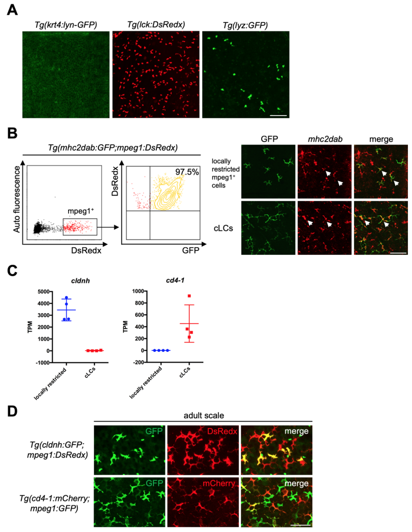
RNA-seq reveals common and unique cell markers for locally restricted mpeg1+ cells and cLCs. Related to Figure 2 (A) Transgenic lines Tg(krt4:lyn-GFP), Tg(lck:DsRedx) and Tg(lyz:GFP) specifically mark keratinocytes, T cells and neutrophils in epidermis respectively. Scale bar, 50μm. (B) Flow cytometry (left) shows 97.5% mpeg1+ cells in epidermis are positive for mhc2dab in Tg(mhc2dab:GFP;mpeg1:DsRedx) fish (n=3). In-situ hybridization of mhc2dab (red) and anti-GFP staining (green) indicate that both locally restricted mpeg1+ cells (green) and cLCs (green) express mhc2dab. White arrows indicate mhc2dab+GFP+ cells. (C) TPM values of cldnh and cd4-1 in locally restricted mpeg1+ cells (blue) and cLCs (red) from four independent RNA-seq data. Each data contains cells isolated from 3 fish. (D) Locally restricted mpeg1+ cells (GFP+DsRedx+, yellow) and cLCs (mCherry+GFP+, yellow) are specifically marked in the epidermis of adult Tg(cldnh:GFP;mpeg1:DsRedx) and Tg(cd4-1:mCherry;mpeg1:GFP) fish respectively (each group, n=8 fish). Scale bar, 50μm.
Reprinted from Developmental Cell, 49(4), Lin, X., Zhou, Q., Zhao, C., Lin, G., Xu, J., Wen, Z., An Ectoderm-Derived Myeloid-like Cell Population Functions as Antigen Transporters for Langerhans Cells in Zebrafish Epidermis, 605-617.e5, Copyright (2019) with permission from Elsevier. Full text @ Dev. Cell