
|
Fig. 1
Zebrafish Epidermis Consists of Two Distinct mpeg1+ Cell Populations
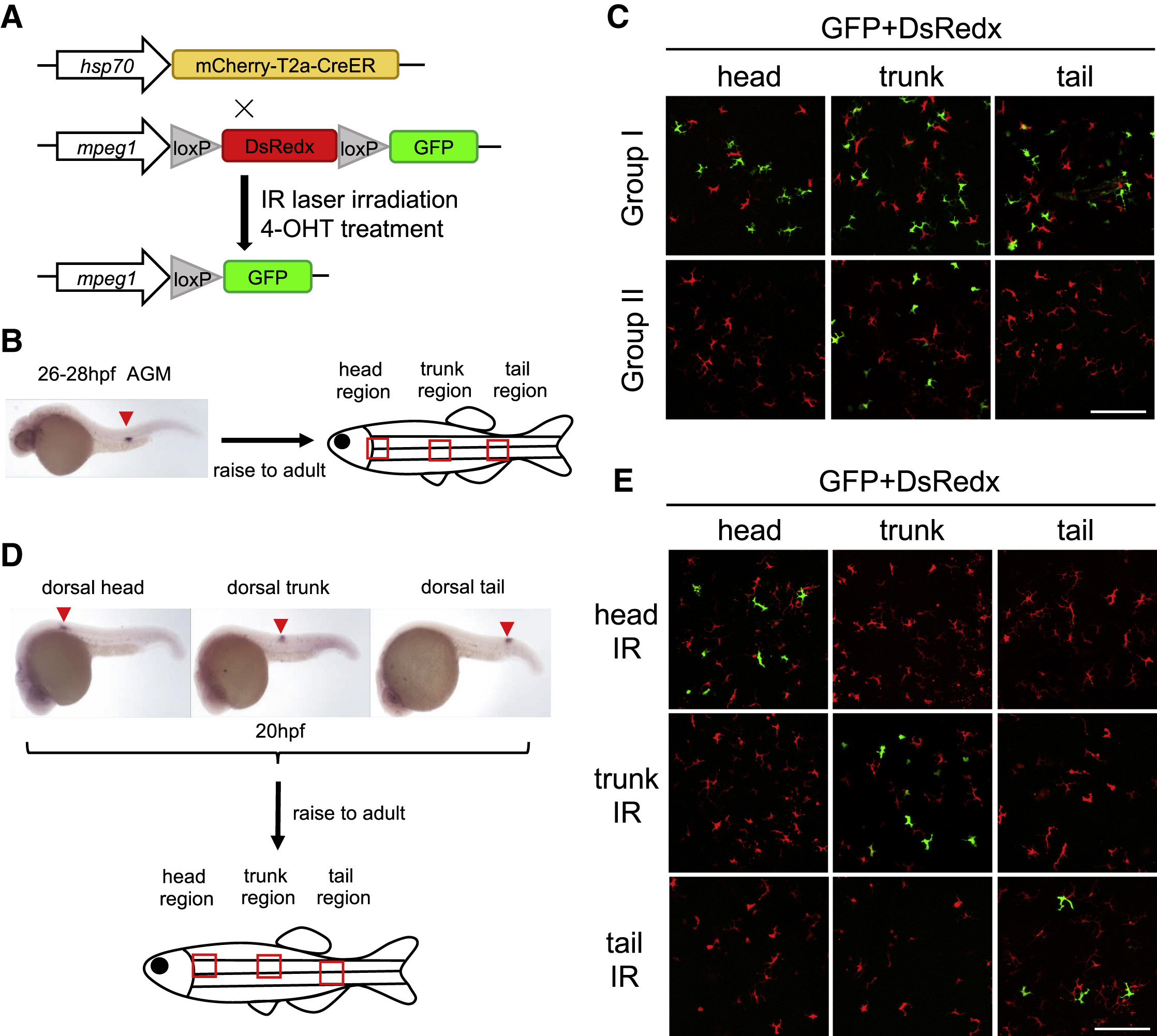
(A) A schematic diagram of IR-LEGO labeling system using infrared and double transgenic line Tg(hsp70:mCherry-T2a-CreERT2;mpeg1:loxP-DsRedx-loxP-GFP). The hsp70 promoter-directed CreER expression is activated in a small region by infrared (IR) laser. After 4-OHT treatment, DsRedx is removed, resulting in GFP expression in macrophage progenies.
(B) A schematic diagram shows the labeling region (expression of CreERT2 mRNA) by 200 MW 1,345 nm infrared laser at 26–28 hpf and the imaging regions (red square) in adult epidermis.
(C) Epidermal GPF+ cells (labeled) and DsRedx+ cells (unlabeled) in the head, trunk, and tail regions in group I (n = 12) and group II (n = 8) adult fish. Scale bar, 100 μm.
(D) In situ hybridization of CreERT2 mRNA expression indicates the labeling positions of non-hematopoietic regions (dorsal head, trunk, and tail) in 20 hpf embryos, and red squares represent the imaging regions.
(E) Epidermal GFP+ cells (labeled) and DsRedx+ cells (unlabeled) in the head, trunk, and tail regions of the adult fish that were irradiated in the dorsal head, trunk, and tail regions at 20 hpf. Scale bar, 100 μm.
Reprinted from Developmental Cell, 49(4), Lin, X., Zhou, Q., Zhao, C., Lin, G., Xu, J., Wen, Z., An Ectoderm-Derived Myeloid-like Cell Population Functions as Antigen Transporters for Langerhans Cells in Zebrafish Epidermis, 605-617.e5, Copyright (2019) with permission from Elsevier. Full text @ Dev. Cell