
|
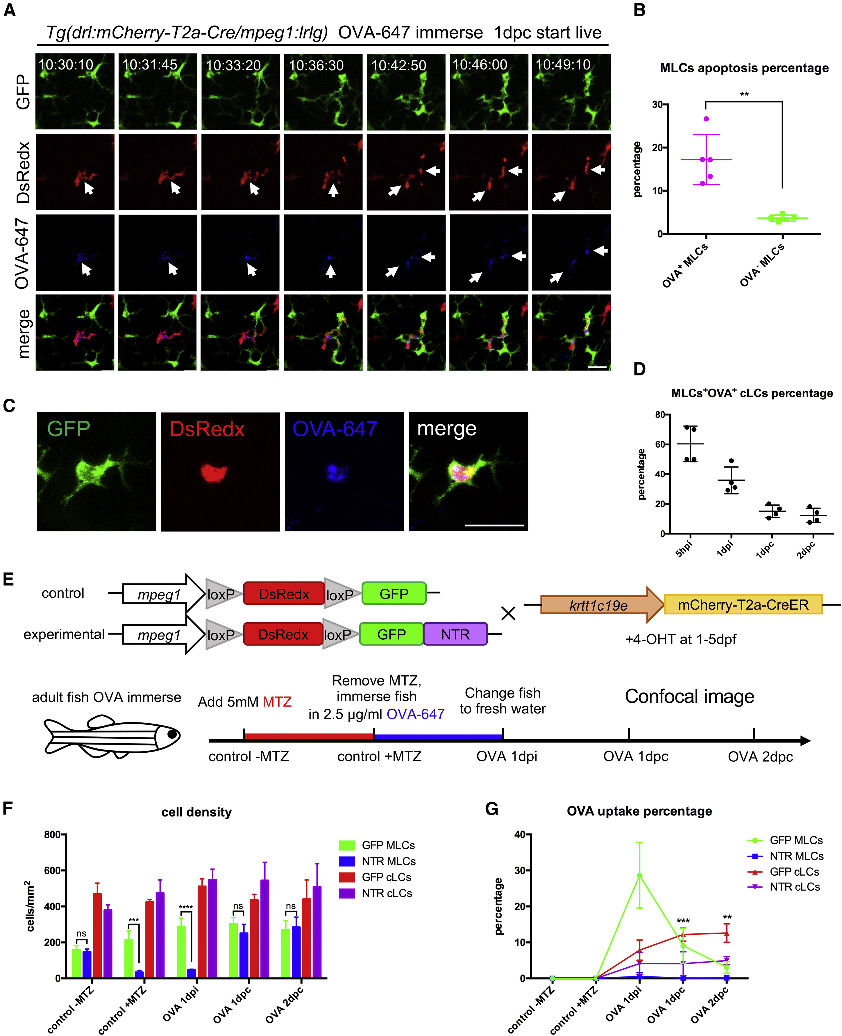
Fig. 6
MLCs Transfer Soluble Antigens to cLCs through Apoptosis-Phagocytosis Axis
(A) Time-lapse imaging on the scales of the OVA-647-treated Tg(drl:mCherry-T2a-Cre;mpeg1:loxP-DsRedx-loxP-GFP) fish. The time-lapse begins at 1 dpc and images for 12 h. White arrows indicate that an apoptotic OVA-647+ (blue) MLC (red), which is subsequently phagocytized by three nearby cLCs (green). Scale bar, 20 μm.
(B) Quantification data show the percentages of apoptotic OVA-647+ and OVA-647- MLCs in the same scales during time-lapse imaging (n = 5 fish).
(C) Images indicate a typical cLCs (green) with OVA-647 (blue) and MLCs debris (red) inside in a living Tg(drl:mCherry-T2a-Cre;mpeg1:loxP-DsRedx-loxP-GFP) fish after OVA-647 treatment. Scale bar, 25 μm.
(D) Quantification data show the percentages of cLCs containing OVA-647 and MLC debris inside to total OVA-647+ cLCs at different time points (n = 4 fish).
(E) A schematic diagram shows the experimental procedure of MLC-depletion strategy using the NTR-MTZ system. Control Tg(krtt1c19e:mCherry-T2a-CreERT2;mpeg1:loxP-DsRedx-loxP-GFP) and experimental Tg(krtt1c19e:mCherry-T2a-CreERT2;mpeg1:loxP-DsRedx-loxP-GFP-NTR) fish (both are treated with 4-OHT for 5 days during early stage) are immersed in 5 mM MTZ for 24 h transferred to system water containing 2.5 μg/mL OVA-647 for another 24 h (OVA 1 dpi), and finally switched to fresh system water.
(F) Quantification data show cell densities of MLCs and cLCs in control (GFP MLCs; GFP cLCs) and experimental fish (NTR MLCs; NTR cLCs) at different time points (each group, n = 4 fish).
(G) Quantification of the percentages of OVA-647+ MLCs and cLCs in control (GFP MLCs; GFP cLCs) and experimental fish (NTR MLCs; NTR cLCs) (each group, n = 4 fish).
Data are represented as mean ± SD, ∗∗p < 0.01, ∗∗∗p < 0.001, ∗∗∗∗p < 0.0001.
Reprinted from Developmental Cell, 49(4), Lin, X., Zhou, Q., Zhao, C., Lin, G., Xu, J., Wen, Z., An Ectoderm-Derived Myeloid-like Cell Population Functions as Antigen Transporters for Langerhans Cells in Zebrafish Epidermis, 605-617.e5, Copyright (2019) with permission from Elsevier. Full text @ Dev. Cell