
|
Fig. S4
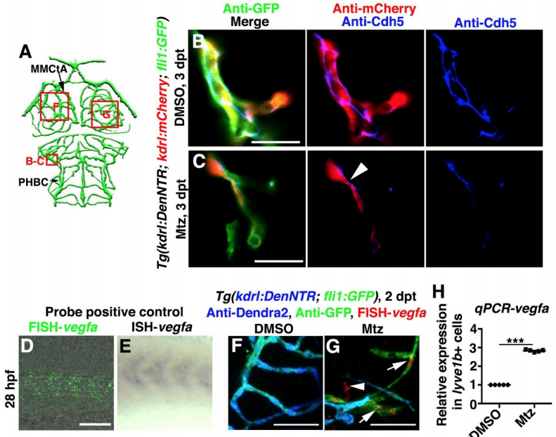
Enrichment of Cdh5 at the lymphatic-vascular interface and expression of vegfa in LECs, Related to Figure 5. (A) Illustrations of lower middle layer brain vascular network indicate the image areas (red boxes) in panels. (B–C) Cdh5 was present in normal cerebrovascular endothelial-endothelial junctions in the control (B, n=16/17). After Mtz treatment, Cdh5 was enriched at the lymphatic (fli1+kdrl-)-vascular (kdrl+) interface at 3 dpt (C, arrowhead, n=14/17). Scale bar, 20 μm. Ventral view, anterior upward. (D–H) The vegfa probe was positive controlled by the comparison of FISH (D, n=21/21) and ISH (E, n=20/20). Lateral view, anterior left. In contrast to the control (F, n=21/22), vegfa expression at 2 dpt was observed in the ingrown LECs (arrows) and other cell types (arrowhead) (G, n=17/20). Ventral view, anterior upward. RT-qPCRs using lyve1b+ cells sorted from the brain confirmed the elevated expression level of vegfa in the ingrown LECs comparing to physiological muLECs (H, n=5). Data are represented as mean ± SEM. ***P<0.001. Scale bar, 50 μm. MMCtA, middle mesencephalic central artery; PHBC, primordial hindbrain channel.v
Reprinted from Developmental Cell, 49(5), Chen, J., He, J., Ni, R., Yang, Q., Zhang, Y., Luo, L., Cerebrovascular Injuries Induce Lymphatic Invasion into Brain Parenchyma to Guide Vascular Regeneration in Zebrafish, 697-710.e5, Copyright (2019) with permission from Elsevier. Full text @ Dev. Cell