
|
Fig. S3
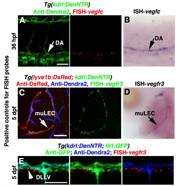
Positive controls for FISH probes, Related to Figure 3. (A and B) The vegfc probe was positive controlled by the comparison of FISH (A, n=23/24) and traditional in situ hybridizations (ISH) (B, n=20/20). Arrows indicate the staining in the dorsal aorta (DA), Lateral view, anterior left. (C–E) The vegfr3 probe was validated by the comparison of FISH (C, n=20/20, dorsal view, anterior upward) and ISH (D, n=21/21, dorsal view, anterior upward) as well as by its staining in the dorsal longitudinal lymphatic vessel (DLLV) (E, arrowhead, n=19/19, lateral view, anterior left). Arrows indicate the staining in the muLECs. Scale bar, 50 μm.
Reprinted from Developmental Cell, 49(5), Chen, J., He, J., Ni, R., Yang, Q., Zhang, Y., Luo, L., Cerebrovascular Injuries Induce Lymphatic Invasion into Brain Parenchyma to Guide Vascular Regeneration in Zebrafish, 697-710.e5, Copyright (2019) with permission from Elsevier. Full text @ Dev. Cell