
|
Fig. S1
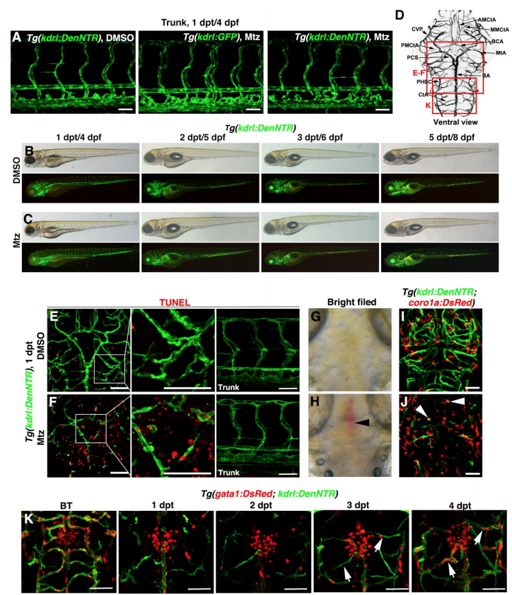
Effects of the Mtz treatment, Related to Figure 1. (A) Mtz treatment was ineffective to the trunk vasculature. (B and C) Compared to DMSO treatment (B, n=20/20), the body phenotypes of larvae treated with Mtz were normal from 1 dpt to 5 dpt (C, n=20/20). Lateral view, anterior left. (D) Boxes on the brain vasculature map indicate image areas in corresponding panels. (E and F) In contrast to DMSO treatment (E, n=32/32), widespread TUNEL signals were detected in the injured brain area including endothelial and neuronal tissues at 1 dpt after Mtz treatment, but not in the trunk vasculature (F, n=29/30). Scale bar, 50 μm. Ventral view, anterior upward. (G and H) In contrast to DMSO treatment (G, n=71/71), Mtz treatment led to hemorrhages (arrowhead) in the injured area (H, n=41/62) at 1 dpt. Dorsal view, anterior upward. (I and J) In contrast to DMSO treatment (I, n=23/23), macrophage infiltration and engulfment of dead cells (arrowheads) were observed after Mtz treatment (J, n=32/32). Dorsal view, anterior upward. (K) Perfusions of nascent blood vessels were initially detectable at 4 dpt under the Tg(gata1:DsRed; kdrl:DenNTR) transgenic background (arrows, n=10/10). Dorsal view, anterior upward. Scale bar, 50 μm. AMCtA, anterior (rostral) mesencephalic central artery; BA, basilar artery; BCA, basal communicating artery; CtA, central artery; CVP, choroidal vascular plexus; MMCtA, middle mesencephalic central artery; MtA, metencephalic artery; PCS, posterior (caudal) communicating segment; PHBC, primordial hindbrain channel; PMCtA, posterior (caudal) mesencephalic central artery.
Reprinted from Developmental Cell, 49(5), Chen, J., He, J., Ni, R., Yang, Q., Zhang, Y., Luo, L., Cerebrovascular Injuries Induce Lymphatic Invasion into Brain Parenchyma to Guide Vascular Regeneration in Zebrafish, 697-710.e5, Copyright (2019) with permission from Elsevier. Full text @ Dev. Cell