
|
Fig. 1
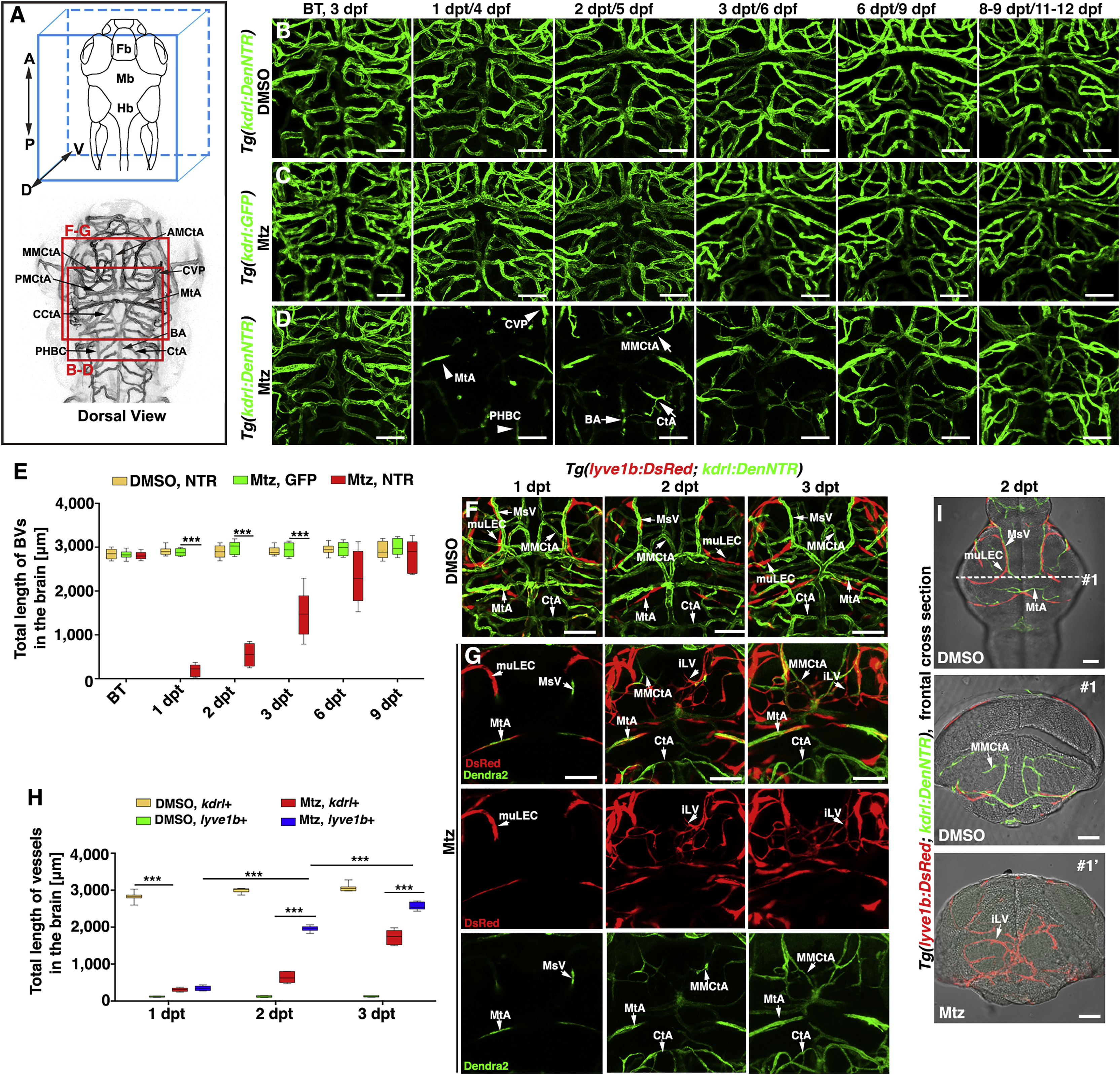
Lymphatic Vessels Appear in the Brain Parenchyma after Cerebrovascular Injury
(A) Brain vascular anatomy of zebrafish at 3 dpf. Red frames indicate the image areas of corresponding panels. Dorsal view, anterior upward.
(B–E) Zebrafish NTR-Mtz cerebrovascular regeneration model. In contrast to the Tg(kdrl:DenNTR) larvae treated with DMSO (B) (n = 40/40) and Tg(kdrl:GFP) larvae treated with Mtz (C) (n = 49/49), brain vasculature of the Tg(kdrl:DenNTR) larvae were specifically injured after incubation with 1 mM Mtz (D) (1 dpt, n = 39/39). A time course of regeneration from BT to 8–9 dpt is shown. The speed of the neoangiogenesis is presented as the total length of the blood vessels (E) (n = 10). Two-way ANOVA by Dunnett’s multiple comparisons test. All p < 0.0001). Arrowheads indicate the remaining relatively large vessels CVP, MtA, and PHBC. Arrows indicate the emerging nascent blood vessels MMCtA, BA, and CtA. ∗∗∗p < 0.001. Data are represented as mean ± SEM. Scale bar, 50 μm. See also Figures S1 and S5.
(F–H) In the control, the lyve1b+ LECs were absent in the brain parenchyma, only present on the optic tectum (F) (n = 24/24) from 1 to 3 dpt. After Mtz treatment (G) (n = 20/24), the growth of the lyve1b+ lymphatics was rapid and active at 2 dpt but apparently slowed at 3 dpt. The quantifications show the total length of kdrl+ and lyve1b+ vessels in the brain at 1–3 dpt (H) (n = 10). Two-way ANOVA by Dunnett’s multiple comparisons test. All p < 0.0001). ∗∗∗p<0.001. Data are represented as mean ± SEM. Scale bar, 50 μm. See also Figure S2.
(I) In frontal cross sections, the lyve1b+ lymphatics were predominantly localized in the meninges at the brain surface in the control larvae (#1) (n = 6/6), whereas became abundant in the brain parenchyma at 2 dpt after Mtz treatment (#1′) (n = 6/6). Scale bar, 50 μm. See also Figure S2.
AMCtA, anterior (rostral) mesencephalic central artery; BA, basilar artery; CCtA, cerebellar central artery; CtA, central artery; CVP, choroidal vascular plexus; Fb, forebrain; Hb, hindbrain; iLVs, ingrown lymphatic vessels; Mb, midbrain; MMCtA, middle mesencephalic central artery; MsV, mesencephalic vein; MtA, metencephalic artery; muLECs, meningeal mural lymphatic endothelial cells; PHBC, primordial hindbrain channel; PMCtA, posterior (caudal) mesencephalic central artery.
Reprinted from Developmental Cell, 49(5), Chen, J., He, J., Ni, R., Yang, Q., Zhang, Y., Luo, L., Cerebrovascular Injuries Induce Lymphatic Invasion into Brain Parenchyma to Guide Vascular Regeneration in Zebrafish, 697-710.e5, Copyright (2019) with permission from Elsevier. Full text @ Dev. Cell