
|
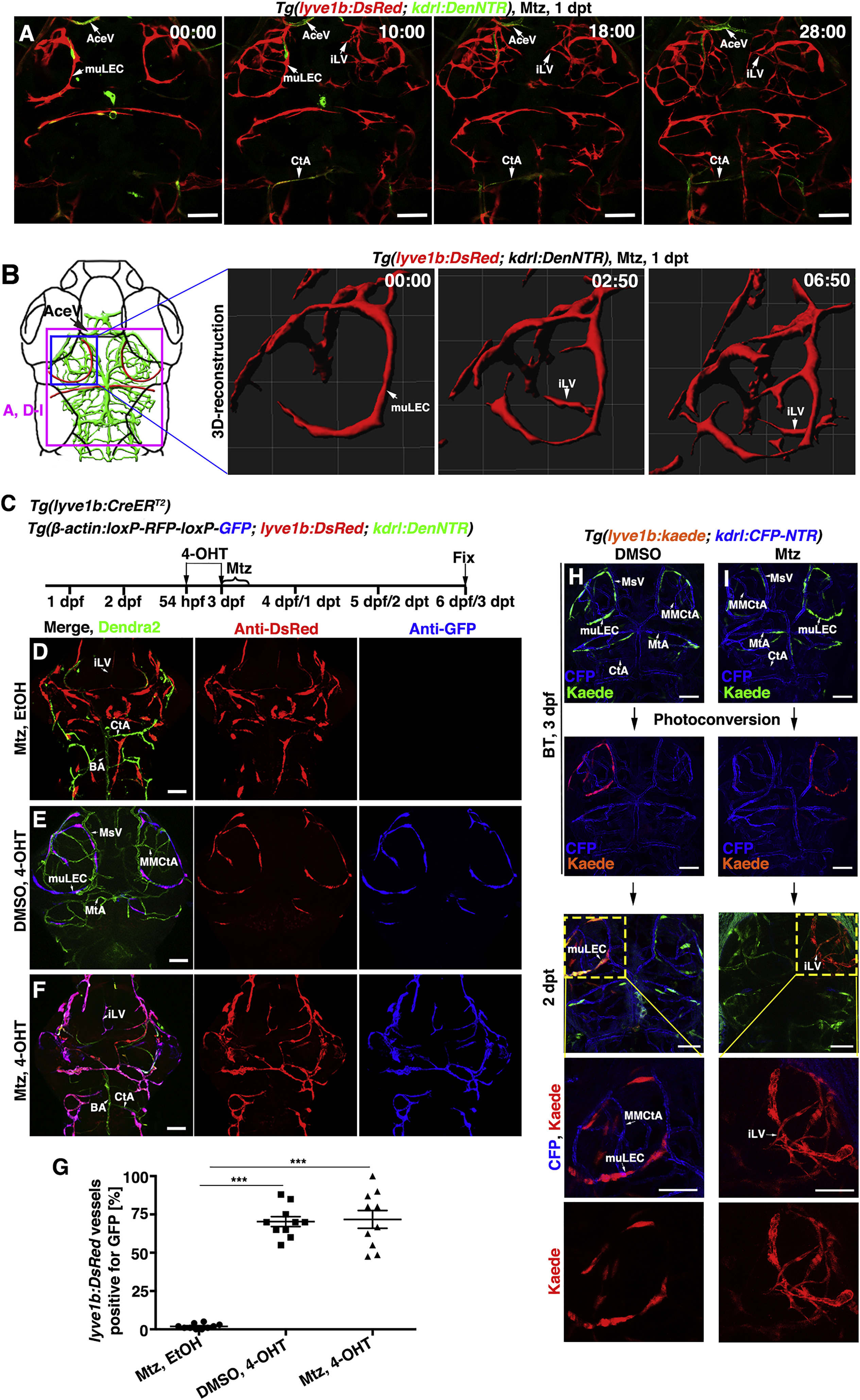
Fig. 2
Meningeal Lymphatics Rapidly Grow into the Injured Parenchyma in Response to Cerebrovascular Damage
(A and B) Time-lapse imaging showed the growth of meningeal lymphatics into the injured area (A) (n = 10/10). Images at the early phases after Mtz treatment showed the initiation of ingrowth and branching of meningeal lymphatics by 3D reconstruction (B) (n = 10/10). The elapsed time is indicated in h:min after 0 dpt. Blue and purple frames indicate the image areas of corresponding panels. See also Videos S1 and S2.
(C–G) The ingrown vessels originate from lyve1b+ cells. Transgenic lines, time points of Mtz and 4-OHT administrations were shown in (C). Negatively controlled by Mtz plus ethanol (D) (n = 12/12) and positively controlled by DMSO plus 4-OHT (E) (n = 19/21), the ingrown vessels were double positive for anti-DsRed and anti-GFP antibodies if Mtz plus 4-OHT were applied (F) (n = 21/24). The statistics show the ratios of the lyve1b:DsRed-labeled vessels positive for GFP (G) (n = 10). Two-tailed unpaired t test, p < 0.0001. ∗∗∗p < 0.001. Data are represented as mean ± SEM. Scale bar, 50 μm.
(H and I) In contrast to DMSO treatment (H) (n = 10/10), photoconversion of muLECs at BT followed by Mtz treatment resulted in retention of Kaede-red fluorescence in the ingrown vessels at 2 dpt (I) (n = 12/12). BT, before treatment. Scale bar, 50 μm.
All images are dorsal view, anterior upward.
AceV, anterior (rostral) cerebral vein; BA, basilar artery; CtA, central artery; iLVs, ingrown lymphatic vessels; MsV, mesencephalic vein; MMCtA, middle mesencephalic central artery; MtA, metencephalic artery; muLECs, meningeal mural lymphatic endothelial cells.
Reprinted from Developmental Cell, 49(5), Chen, J., He, J., Ni, R., Yang, Q., Zhang, Y., Luo, L., Cerebrovascular Injuries Induce Lymphatic Invasion into Brain Parenchyma to Guide Vascular Regeneration in Zebrafish, 697-710.e5, Copyright (2019) with permission from Elsevier. Full text @ Dev. Cell