
|
Fig. 4
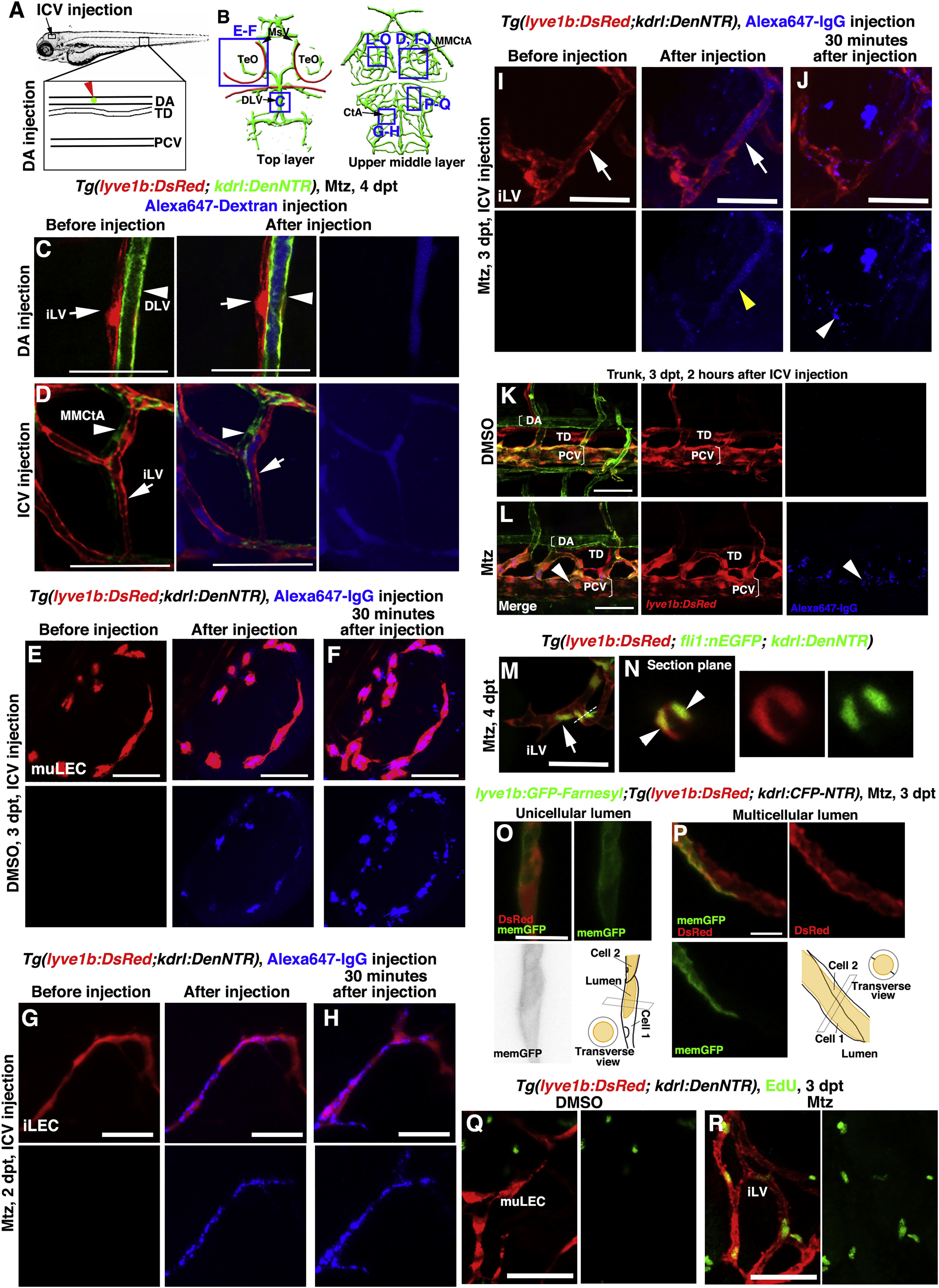
The Ingrown Lymphatics Undergo Lumenization to Drain Brain Interstitial Fluid
(A–D) The ingrown meningeal lymphatics become lumenized to drain interstitial fluid. Illustrations of dorsal aorta (DA) and ICV (intracerebroventricular) injection points (A). Blue frames indicate the image areas in the corresponding panels (B). Alexa647-dextran was filled in the blood vessels (arrowheads) but remained absent in the meningeal lymphatics (arrows) after DA injection (C) (n = 9/11). Drainage of Alexa647-labeled interstitial fluid was rapidly observed in the ingrown lymphatic vessels (arrows) after ICV injection (D) (n = 16/18). Note that the lumen of the lymphatic vessel was visible (D, arrows). Dorsal view, anterior upward. Scale bar, 50 μm.
(E–L) Endocytosis, but not lumen flow of Alexa647-IgG, was observed in the muLECs of the control larvae after ICV injection at 3 dpt (E) (n = 15/15) and was also observed in the ingrown LECs of the Mtz-treated larvae after ICV injection at 2 dpt (G) (n = 12/13). 30 min later, the endocytosed dye maintained and accumulated in these LECs (F) (n = 14/14; (H) (n = 11/12). If the Alexa647-IgG was injected at 3 dpt after Mtz treatment, the dye filled in the lumen of lymphatic vessels immediately after injection (I) (yellow arrowhead) (n = 17/20). 30 min later, the dye was drained out from the lumen, but the endocytosed dye remained in the ingrown LECs (J,) (arrowheads) (n = 16/20). Comparing to the non-injured larvae (K) (n = 18/18), the dye became apparent in the PCVs of injured larvae at 2 h post-injection (L) (arrowheads) (n = 17/18). Note that the lumen of lymphatics was clearly visible at 3 dpt (I) (arrows) but not in the control (E) or at 2 dpt (G). Dorsal view, anterior upward. Scale bar, 50 μm.
(M and N) The nuclei of LECs are labeled by nuclear EGFP transgenes. The section plane in (M) (dash line) is presented in (N). Arrow and arrowheads indicate unicellular and multicellular lumens, respectively. n = 11/12. Dorsal view, anterior upward. Scale bar, 20 μm.
(O and P) The plasma membrane of LECs is mosaically labeled by the injection of membrane GFP plasmid. The unicellular lumen is presented by membrane GPF epifluorescence (O) (n = 22/24), whereas the multicellular lumen is exhibited by the mosaicity of a single vessel (P) (n = 22/24), both with schematic illustrations. Scale bar, 10 μm.
(Q and R) In contrast to the muLECs in the control (Q) (n = 10/10), many of the ingrown LECs were positive for EdU labeling at 3 dpt (R) (n = 12/12). Ventral view, anterior upward. Scale bar, 20 μm.
CtA, central artery; DA, dorsal aorta; DLV, dorsal longitudinal vein; iLVs, ingrown lymphatic vessels; MMCtA, middle mesencephalic central artery; MsV, mesencephalic vein; muLECs, meningeal mural lymphatic endothelial cells; PCV, posterior cardinal vein; TD, thoracic duct; TeO, optic tectum.
Reprinted from Developmental Cell, 49(5), Chen, J., He, J., Ni, R., Yang, Q., Zhang, Y., Luo, L., Cerebrovascular Injuries Induce Lymphatic Invasion into Brain Parenchyma to Guide Vascular Regeneration in Zebrafish, 697-710.e5, Copyright (2019) with permission from Elsevier. Full text @ Dev. Cell