
|
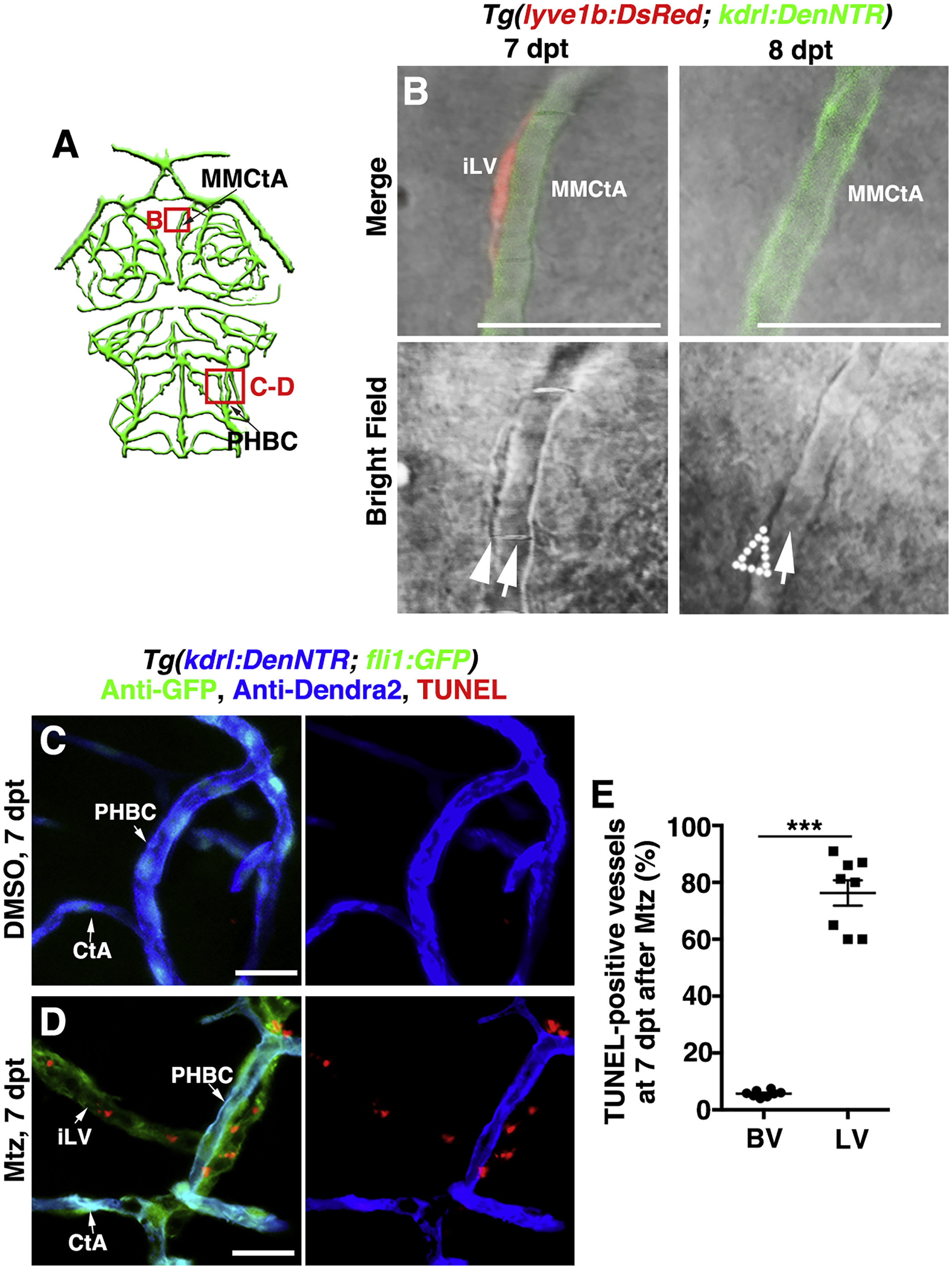
Fig. 6
The Ingrown Lymphatic Vessels Undergo Apoptosis after the Completion of Cerebrovascular Regeneration
(A) Illustrations of brain vasculature indicate the image areas (red boxes) in panels. All panels are dorsal views, anterior upward.
(B) The ingrown lymphatic vessels disappeared from 7 to 8 dpt (n = 23/26). Arrowheadand arrows indicate the adhering lymphatic and blood vessels, respectively. Note the disappearance of the lymphatic vessel at 8 dpt (dotted arrowhead). Scale bar, 20 μm.
(C–E) In contrast to the control (C) (n = 33/35), TUNEL signals were detected in approximately 70% of the ingrown lymphatic vessels (fli1+kdrl−) (D) (n = 30/33) but in less than 5% of the blood vessels (kdrl+) (E) (n = 8). Two-tailed unpaired t test (p < 0.0001). Scale bar, 20 μm. Data are represented as Mean ± s.e.m., ∗∗p < 0.001. See also Figure S6.
CtA, central artery; iLV, ingrown lymphatic vessels; MMCtA, middle mesencephalic central artery; PHBC, primordial hindbrain channel.
Reprinted from Developmental Cell, 49(5), Chen, J., He, J., Ni, R., Yang, Q., Zhang, Y., Luo, L., Cerebrovascular Injuries Induce Lymphatic Invasion into Brain Parenchyma to Guide Vascular Regeneration in Zebrafish, 697-710.e5, Copyright (2019) with permission from Elsevier. Full text @ Dev. Cell