
|
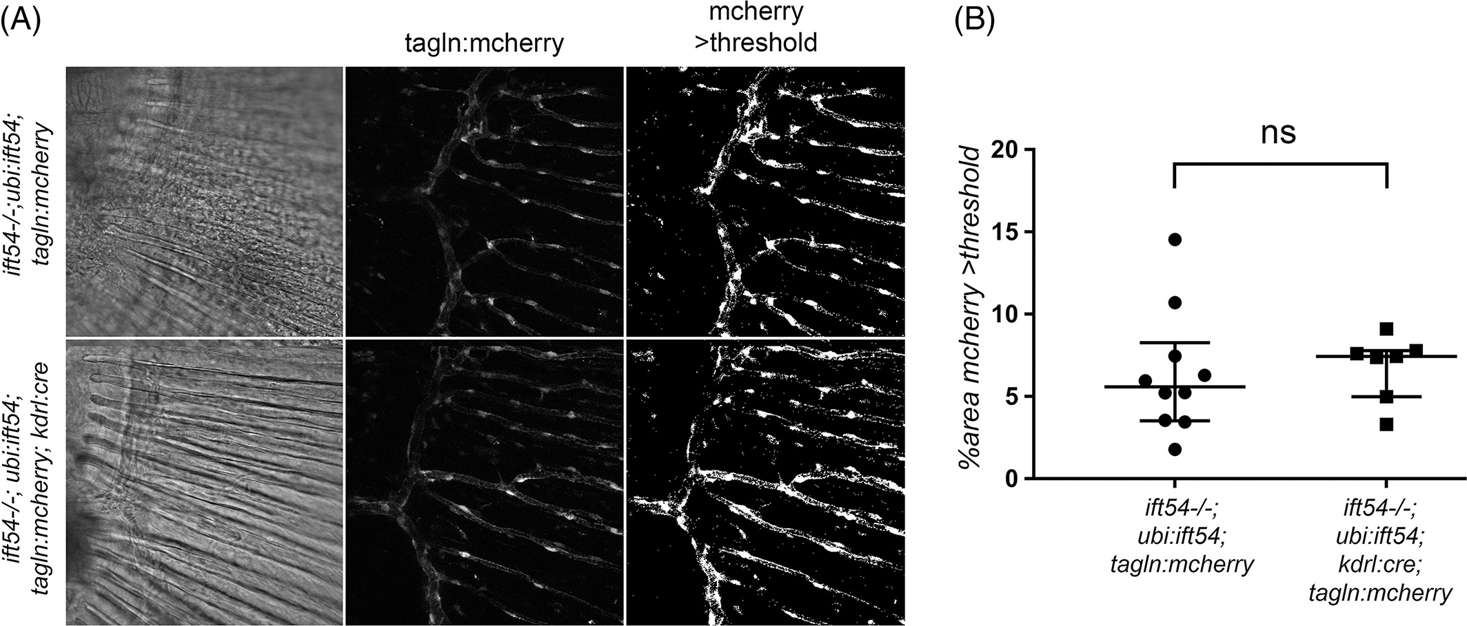
Fig. 12
Endothelial‐specific excision of the Tg(ubi:loxP‐ift54‐loxP‐myr‐mcherry,myl7:EGFP)sh488‐rescuing transgene causes no apparent further impairment to perivascular mural cells in the caudal fin vascular plexus of ift54 tp49; Tg(ubi:loxP‐ift54‐loxP‐myr‐mcherry,myl7:EGFP)sh488 fish. A: Confocal stack projections showing mcherry fluorescence from 17‐dpf caudal fins of ift54tp4; Tg(ubi:loxP‐ift54‐loxP‐myr‐mcherry,myl7:EGFP)sh488 /+; Tg(tagln:mcherry)sh441 /+ (ift54−/−; ubi:ift54; tagln:mcherry) and ift54 tp4; Tg(ubi:loxP‐ift54‐loxP‐myr‐mcherry,myl7:EGFP)sh488/+;Tg(kdrl:cre)s898; Tg(tagln:mcherry)sh441 /+ (ift54−/−; ubi:ift54; tagln:mcherry; kdrl:cre). Also shown are binary threshold processed images as used for quantification of the area of Tg(tagln:mcherry)sh441 /+ expression. Single‐focal‐plane transmitted light views are shown for orientation. B: Chart shows % of field of view with >threshold mcherry expression as measured from processed confocal projections of caudal fins. Overlay shows median and interquartile range. Two‐tailed Mann‐Whitney test P = 0.5362. The Tg(kdrl:cre)s898 /+ caused no apparent impairment to mural cell coverage in the newly developed fin vascular bed in the fish with partial transgenic rescue of ift54 tp49