
Image
|
Figure Caption
Fig. S3
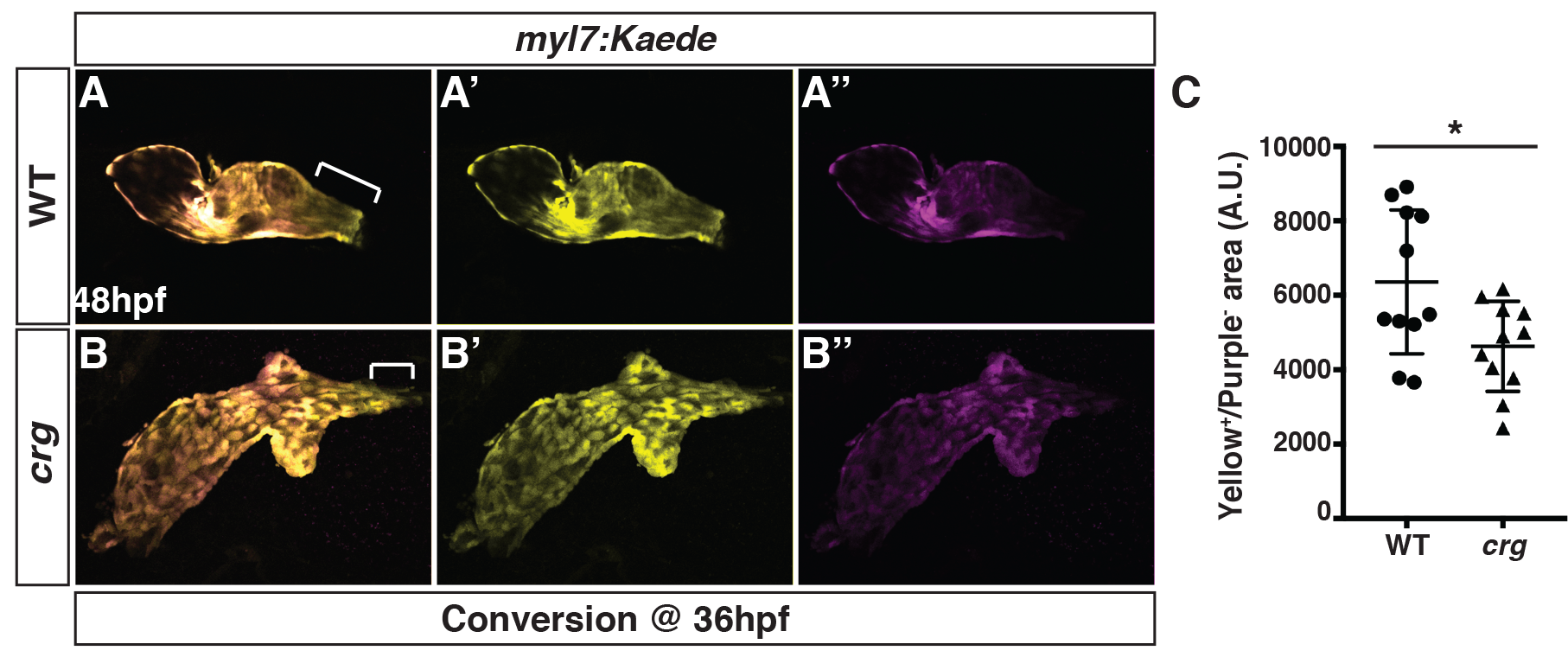
Later-differentiating VCs are reduced in crg mutants.
(A-B”) Images of hearts from photoconverted WT sibling and crg mutant myl7:Kaede embryos at 48 hpf following photoconversion at 36 hpf. The arterial poles (brackets) are to the right. (C) Quantification of the area of later-differentiating VCs (Yellow+/Purple-). (n = 11 for WT and crgmutants).
Acknowledgments
This image is the copyrighted work of the attributed author or publisher, and
ZFIN has permission only to display this image to its users.
Additional permissions should be obtained from the applicable author or publisher of the image.
Full text @ PLoS Genet.