
Image
|
Figure Caption
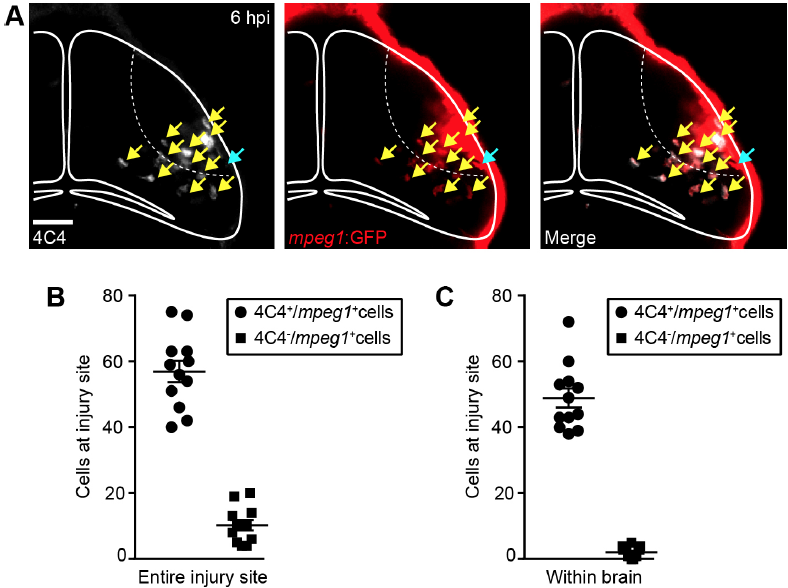
Fig. S9
The majority of mpeg1+ cells at the injury site within the brain are also labelled by 4C4 immunohistochemistry. (A) Confocal images of the optic tectum at 6 hpi in mpeg1:GFP animals after 4C4 and GFP immunohistochemistry. Yellow arrows indicate 4C4+/mpeg1+ cells. Light blue arrow indicates a 4C4-/mpeg1+ cell. Scale bar, 40 μm. (B,C) Quantification of 4C4+/mpeg1+ and 4C4-/mpeg1+ cells at the entire injury site (B) or within the brain (C) at 6 hpi. n = 12 animals
Acknowledgments
This image is the copyrighted work of the attributed author or publisher, and
ZFIN has permission only to display this image to its users.
Additional permissions should be obtained from the applicable author or publisher of the image.
Full text @ Development