IMAGE

Fig. 3

ID
ZDB-IMAGE-190801-5
Source
Figures for Nowak-Sliwinska et al., 2018
Image
Figure Caption

Fig. 3

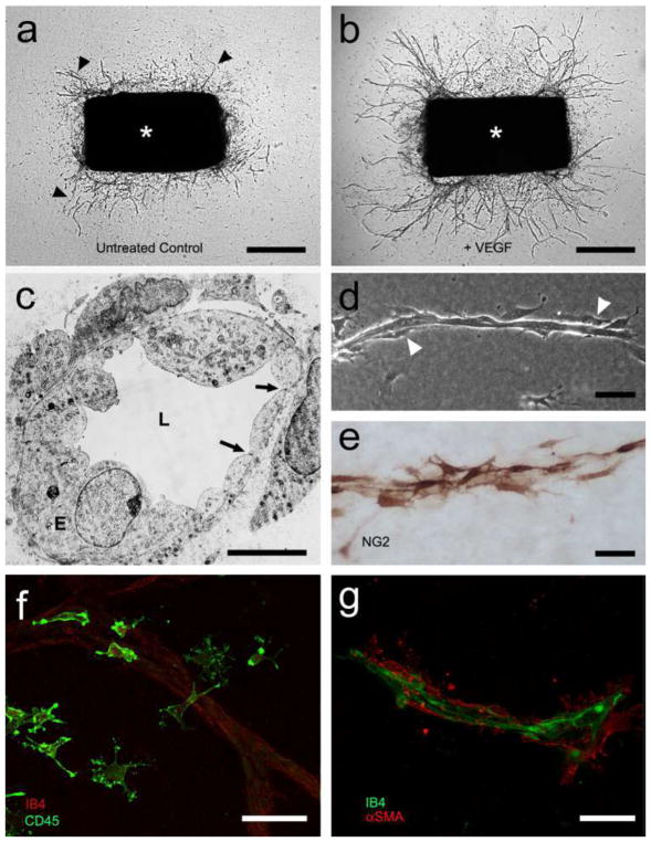
Aortic ring assay of angiogenesis. a Serum-free collagen gel culture of rat aorta (asterisk) photographed at day 6 (microvessels marked by arrowheads). b Aortic culture treated with VEGF (5 ng/ml) shows increased number of microvessels (day 6). c Electron micrograph of aorta-derived microvessel with polarized endothelium (E), patent lumen (L), and surrounding pericytes (P); endothelial tight junctions are marked by arrows. d Phase-contrast micrograph of microvessel composed of an inner core of endothelial cells and surrounding pericytes (white arrowheads). e Pericytes highlighted with immunoperoxidase stain for NG2. f Immunofluorescent image of aorta-derived macrophages stained for CD45; an isolectin B4 (IB4)-stained endothelial sprout is visible in the background. g Confocal image of microvessel double stained for endothelial cells (IB4) and pericytes (alpha smooth muscle actin, αSMA). Magnification bars = 500 μm (a, b), 5 μm (c), 50 μm (dg)

Acknowledgments
This image is the copyrighted work of the attributed author or publisher, and ZFIN has permission only to display this image to its users. Additional permissions should be obtained from the applicable author or publisher of the image. Full text @ Angiogenesis