IMAGE

Fig. 2

ID
ZDB-IMAGE-190801-4
Source
Figures for Nowak-Sliwinska et al., 2018
Image
Figure Caption

Fig. 2

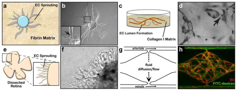
Three-dimensional assays of vascular morphogenesis. a A fibrin bead assay uses collagen I- and EC-coated Cytodex beads embedded within a 3D fibrin gel matrix to measure EC sprouting and lumen formation. b These features are readily resolved using phase-contrast microscopy. c EC tube formation can be measured by embedding EC within a collagen I matrix. d Once formed, these tubes can be visualized by toluidine blue staining and bright-field microscopy. e Whole-mount, dissected retinas from postnatal mice are mounted within collagen I-Matrigel matrix mix and cultured in pro-angiogenic medium to stimulate EC sprouting. f Sprout and lumen formation are resolved using phase-contrast microscopy. g The vascularized micro-organ (VMO) approach utilizes “arteriole” (high pressure) and “venule” (low pressure) microfluidic channels to drive medium diffusion and flow across a cell chamber where microvasculature forms. h The formed microvasculature (EC, red) can be measured for leak by perfusion with 70 kDa FITC-dextran (green)

Acknowledgments
This image is the copyrighted work of the attributed author or publisher, and ZFIN has permission only to display this image to its users. Additional permissions should be obtained from the applicable author or publisher of the image. Full text @ Angiogenesis