IMAGE

Fig. 21

ID
ZDB-IMAGE-190801-23
Source
Figures for Nowak-Sliwinska et al., 2018
Image
Figure Caption

Fig. 21

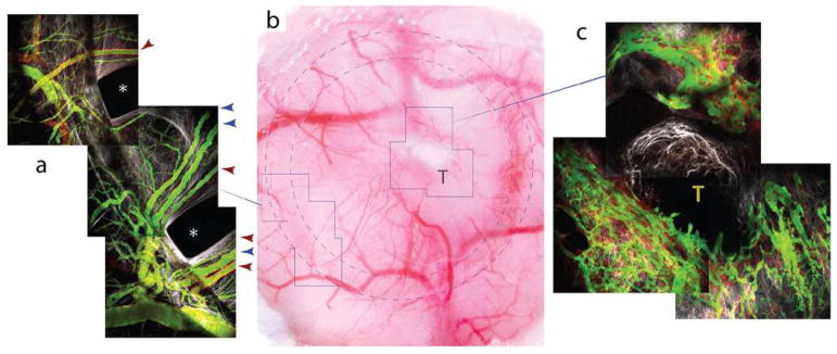
MMTV tumor vasculature in the cranial window pillar TIC. a MPLSM/SHG image of the indicated region in the bright-field image (b). Near the edge of the PDMS, the vasculature extends radially into the central chamber. At this time point (day 7 after implantation), the vasculature is mature and has normal morphology in the regions far from the tumor. Four feeding arterioles (red arrowheads) and three venules (blue arrowheads) are indicated. These vessels have significant flow and have acquired smooth muscle cells in their walls (red, αSMA+-DsRed). Note that the arterioles generally have more αSMA signal than the venules, as expected. c The vasculature near the growing tumor has dramatically different morphology and flow, as observed in other animal models and human tumors. The tumor was not fluorescently labeled in this group, but is visible as the mass extending from the central tumor (T) in b (from [493])

Acknowledgments
This image is the copyrighted work of the attributed author or publisher, and ZFIN has permission only to display this image to its users. Additional permissions should be obtained from the applicable author or publisher of the image. Full text @ Angiogenesis