IMAGE

Fig. 13

ID
ZDB-IMAGE-190801-16
Source
Figures for Nowak-Sliwinska et al., 2018
Image
Figure Caption

Fig. 13

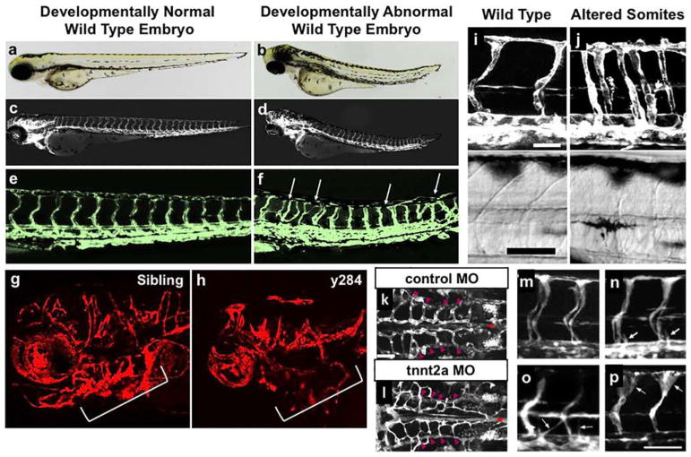
Assessing embryonic morphology and effects on vascular patterning. af Transmitted light (a, b), epifluorescence (c, d), and confocal (e, f) images of three dpf developmentally normal (a, c, e) or developmentally abnormal (b, d, f) embryos from the same Tg(fli1:egfp)y1 transgenic “wild-type” zebrafish population. The normal animal has a normal morphology (a) and normal vessel patterning (c, e), while the developmentally abnormal animal has a stunted, somewhat malformed trunk (B) and also displays trunk intersegmental vessel patterning defects (d, f). g, h Confocal images of the cranial vasculature in 48 hpf wild-type sibling (g) and y284 mutant (h) Tg(kdrl:mRFP-F)y286 animals. The y284 mutants have small heads heads and eyes, accompanied by reduced and abnormal formation of cranial vessels and aortic arches (brackets). The vascular defects should be interpreted with caution, as they can be primary or solely a consequence of the smaller head (or possibly both). Transplantation and/or mosaic transgenic expression can be used to assess the vascular cell autonomy of phenotypes. i, j Confocal (top) and corresponding transmitted light (bottom) images of 48 hpf Tg(fli1:egfp)y1 wild-type sibling (i) and fused somites (fssy66) mutant (j) animals. Improper somite formation in fssy66 mutants (lack of chevron shaped somite segments, seen in the bright-field images) indirectly results in altered intersegmental vessel patterning (j, top). kp Confocal images of the hindbrain vasculature (k, l) or trunk intersegmental vessels (mp) in Tg(fli1:egfp)y1 control (k) or cardiac troponin T type 2a (tnnt2a) (lp) deficient animals at 1.5 (m), 2 (k, l, n), 2.5 (o), or 3.5 (p) days post-fertilization (dpf). Control animals have normal blood flow, but tnnt2a-deficient animals have no heart beat and lack all blood flow. Although formation of the vasculature is largely normal for several days in the absence of blood flow (l, n, o), abnormalities in vessel growth and patterning begin to appear at later stages, such as enlargement of the dorsal intersegmental vessels at 3.5 dpf (p). This illustrates the need to analyze vascular phenotypes as early in development as possible in zebrafish with absent or defective blood flow. All images are lateral views, rostral to the left, except panels k and l, which show dorsal views, rostral to the left. Images in panels i and j are from Ref. [306], images in panels k and l are from Ref. [650], and images in panels mp are from Ref. [651]. Scale bars = 50 μm

Acknowledgments
This image is the copyrighted work of the attributed author or publisher, and ZFIN has permission only to display this image to its users. Additional permissions should be obtained from the applicable author or publisher of the image. Full text @ Angiogenesis