IMAGE

Fig. 8

ID
ZDB-IMAGE-190801-10
Source
Figures for Nowak-Sliwinska et al., 2018
Image
Figure Caption

Fig. 8

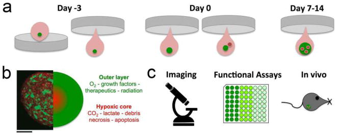
EC co-culture spheroid assay. a Overview of the procedure of the hanging drop spheroid growth assay over time. Prior to initiating the co-culture, tumor cells (green) are allowed to establish initial micro-spheroids by themselves. Typically, after 72 h EC can be introduced (red) and their incorporation monitored over time up to 14 days. b After 7 days, the tumor-EC spheroid can be quantitatively and qualitatively evaluated by various techniques, such as c (fluorescence) microscopy, histology, functional assays or implanted in vivo. Breast tumor cells (green; GFP-4T1) are combined with 2H11 EC (red) stained with MitoTracker Red. Scale bar = 100 μm

Acknowledgments
This image is the copyrighted work of the attributed author or publisher, and ZFIN has permission only to display this image to its users. Additional permissions should be obtained from the applicable author or publisher of the image. Full text @ Angiogenesis