
Image
|
Figure Caption
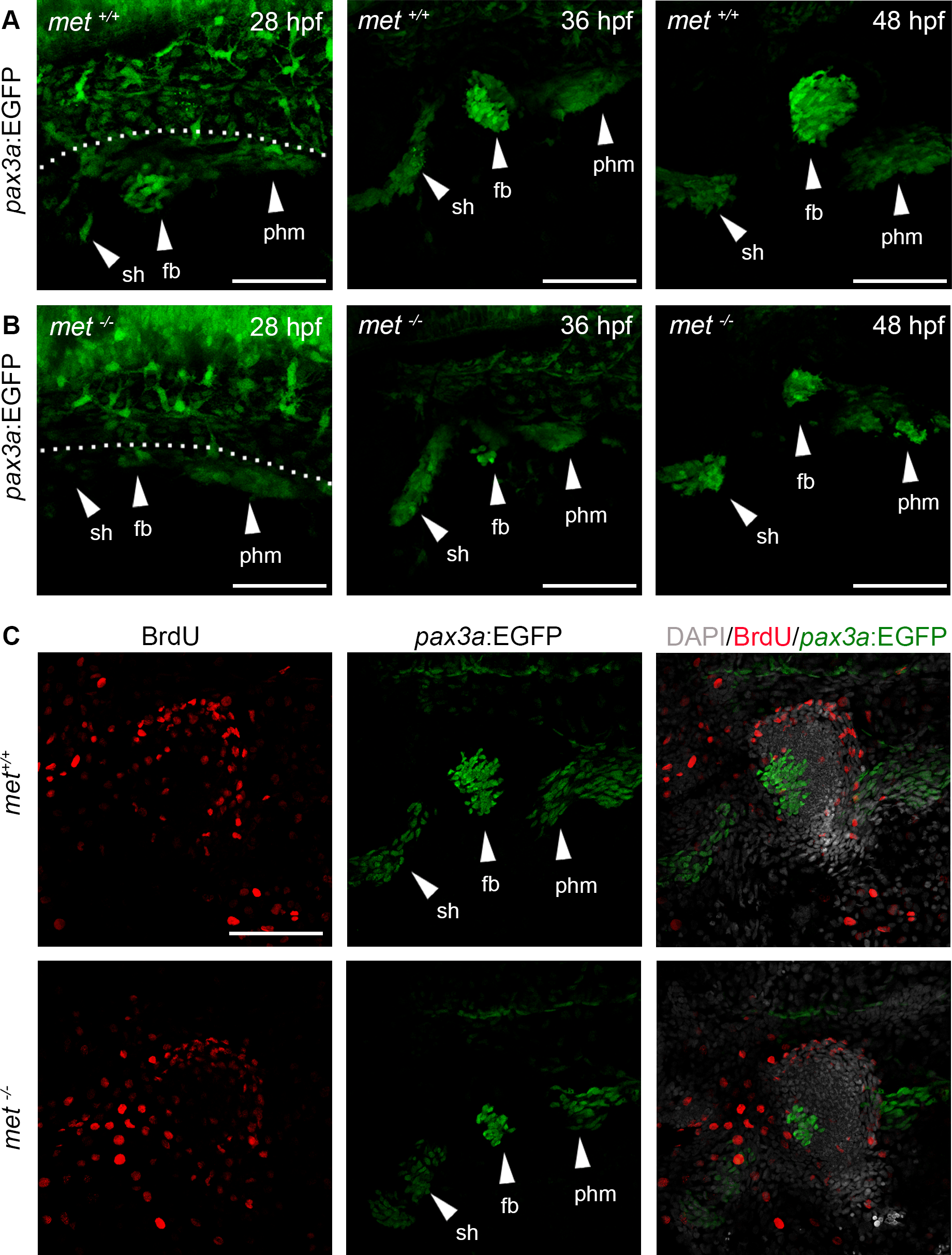
Fig. 6
The pax3a:EGFP+ populations of MMPs migrating out from the somites are reduced in met-/- mutants.
Lateral view of transgenic pax3a:EGFP expression in (A) met+/+ siblings and (B) met-/-mutant embryos at 28 hpf (n = 6 for met+/+ and n = 9 for met-/-), 36 hpf (n = 7 for met+/+and n = 5 for met-/-) and 48 hpf (n = 5 for met+/+ and n = 5 for met-/-). (C) met+/+ siblings (n = 5) and met-/- mutant (n = 5) pax3a:EGFP (green) embryos treated with BrdU (red) from 24 to 48 hpf to visualize proliferating cells. Dashed line in A indicate yolk-somite border. Abbreviations: sh: sternohyoideus; fb: fin bud; phm: posterior hypaxial muscle. Scale bar: 50 μm.
Figure Data
Acknowledgments
This image is the copyrighted work of the attributed author or publisher, and
ZFIN has permission only to display this image to its users.
Additional permissions should be obtained from the applicable author or publisher of the image.
Full text @ PLoS One