
Image
|
Figure Caption
Fig. S6
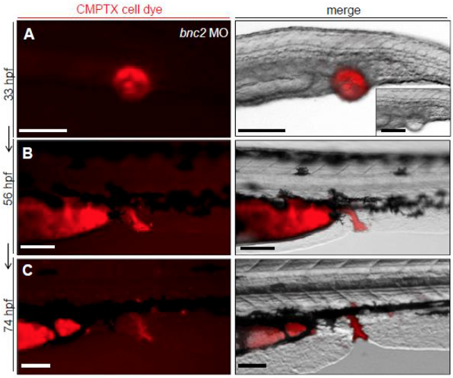
Injection of Fluorescent Cell Dye Tracker CMTPX Indicates Surrounding Cells of Pronephric Outlet Obstruction to Later Form Parts of Distal Pronephric Ducts and the Cloaca
(A-C): Injection of fluorescent cell dye tracker CMPTX into the pronephric outlet obstruction ‘vesicle’ of a bnc2 MO injected zebrafish embryo at 33 hpf and follow-up imaging of the same individual zebrafish demonstrates that cells forming the ‘vesicle’ become part of the distal end of the pronephric ducts and the cloaca later in development (56 and 74 hpf).
Acknowledgments
This image is the copyrighted work of the attributed author or publisher, and
ZFIN has permission only to display this image to its users.
Additional permissions should be obtained from the applicable author or publisher of the image.
Full text @ Am. J. Hum. Genet.