IMAGE

Fig 3

ID
ZDB-IMAGE-190723-833
Genes
Antibodies
Source
Figures for Song et al., 2019
Image
Figure Caption

Fig 3 SHF-derived VC and smooth muscle development is impaired in the <italic>crg</italic> mutants.

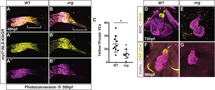
(A-B”) Representative images of hearts from photoconverted WT sibling and crg mutant myl7:NLS-KikGR embryos at 48 hpf. The arterial poles (brackets) are to the right. (C) Quantification of later-differentiating VCs (Yellow+/Purple- cells) (n = 10 for WT and crg mutants). (D,E) Confocal images of IHC for MHC and Elnb in WT and crg mutant embryos at 72 hpf. n = 10 WT and n = 10 crg mutants embryos examined. (F,G) Confocal images of DAF-2DA staining coupled with IHC for Vmhc in WT sibling and crg mutant embryos at 96 hpf. n = 10 WT and n = 10 crg mutants embryos examined. Images in D-G are frontal views with anterior up. Arrows indicate Elnb and DAF-2DA staining of the bulbous arteriosus.

Figure Data
Acknowledgments
This image is the copyrighted work of the attributed author or publisher, and ZFIN has permission only to display this image to its users. Additional permissions should be obtained from the applicable author or publisher of the image. Full text @ PLoS Genet.