IMAGE

Fig 2

ID
ZDB-IMAGE-190723-832
Genes
Source
Figures for Song et al., 2019
Image
Figure Caption

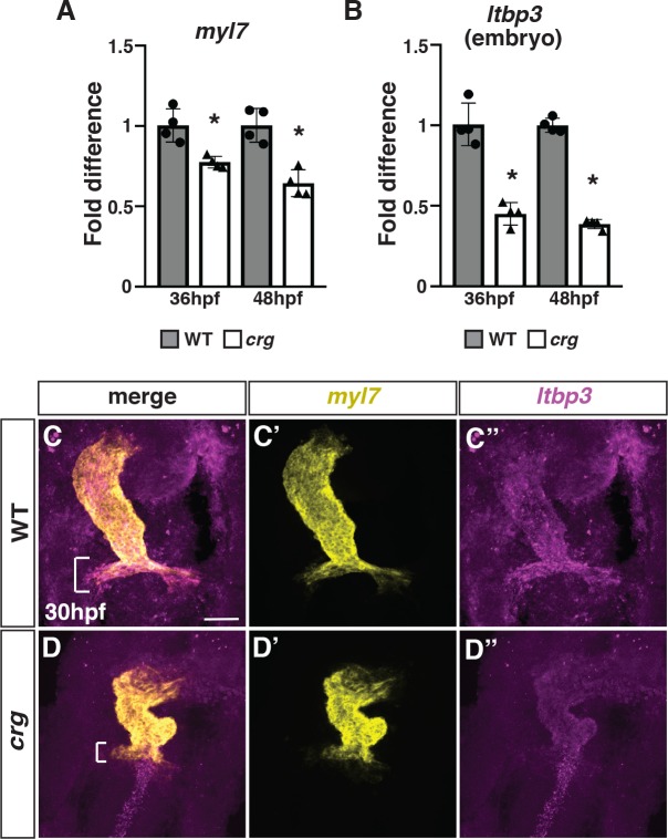
Fig 2 SHF markers of the arterial pole are reduced in <italic>crg</italic> mutants.

(A,B) RT-qPCR for the pan-cardiac differentiation marker myl7 and SHF marker ltbp3 from embryos at 36 and 48 hpf. (C-D”) Two-color FISH for myl7 and ltbp3 in WT sibling and crg mutant embryos at 30 hpf. Brackets in C and D indicate presence and absence of ltbp3 at the arterial pole of WT and crg mutant hearts, respectively. n = 5 WT and n = 5 crg mutants embryos were examined.

Figure Data
Acknowledgments
This image is the copyrighted work of the attributed author or publisher, and ZFIN has permission only to display this image to its users. Additional permissions should be obtained from the applicable author or publisher of the image. Full text @ PLoS Genet.