
|
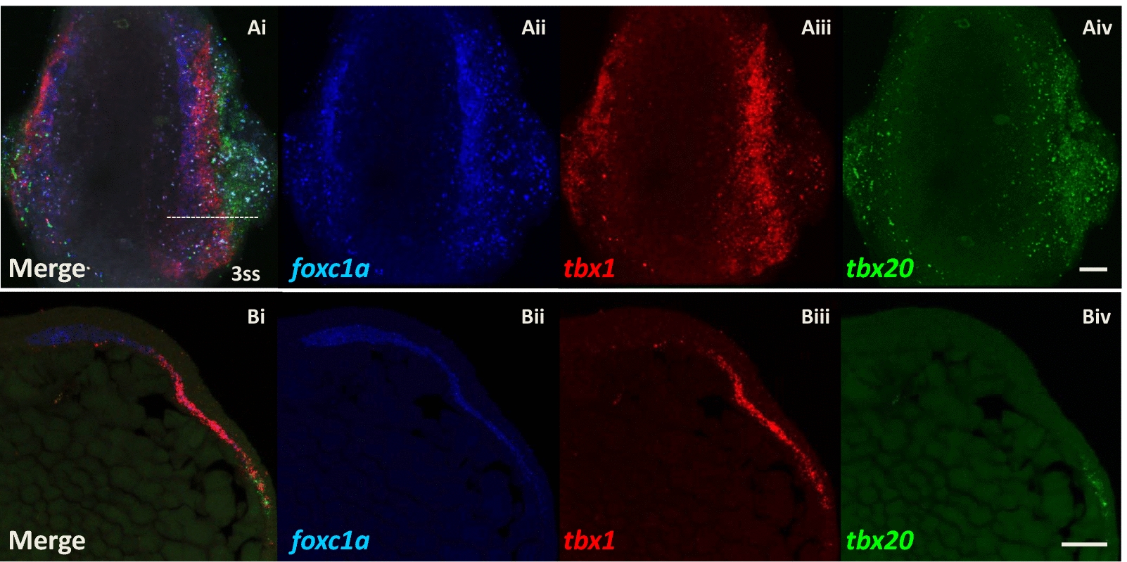
Fig. 5
Expression patterns of foxc1a, tbx1 and tbx20 genes resolve into three longitudinal strips in zebrafish head mesoderm (3ss, 11hpf). Confocal microscope images of triple in situ hybridisation of foxc1a (dark blue), tbx1 (red) and tbx20 (green) mRNA. Ai–iv Dorsal views of a whole-mount zebrafish embryo at 3ss (11hpf), anterior is oriented to the top. Note that the embryo was slightly tilted towards to the left when embedded in agarose gel to show better the separation of expression regions on the right side of the embryo. The innermost paraxial strip (dark blue) expresses foxc1a gene, then a more lateral strip (red) expresses tbx1, and finally, the most lateral strip (green) expresses tbx20. The boundaries of the three regions overlap. Bi–iv Cross sections of a zebrafish embryo at 3ss showing the right side of the head mesoderm, dorsal is oriented to the left-top. The level of sectioning is at the anterior hindbrain shown as the dotted line in Ai. Scale bar: 50 μm