IMAGE

Fig. 5

ID
ZDB-IMAGE-190723-762
Source
Figures for Kimura et al., 2019
Image
Figure Caption

Fig. 5

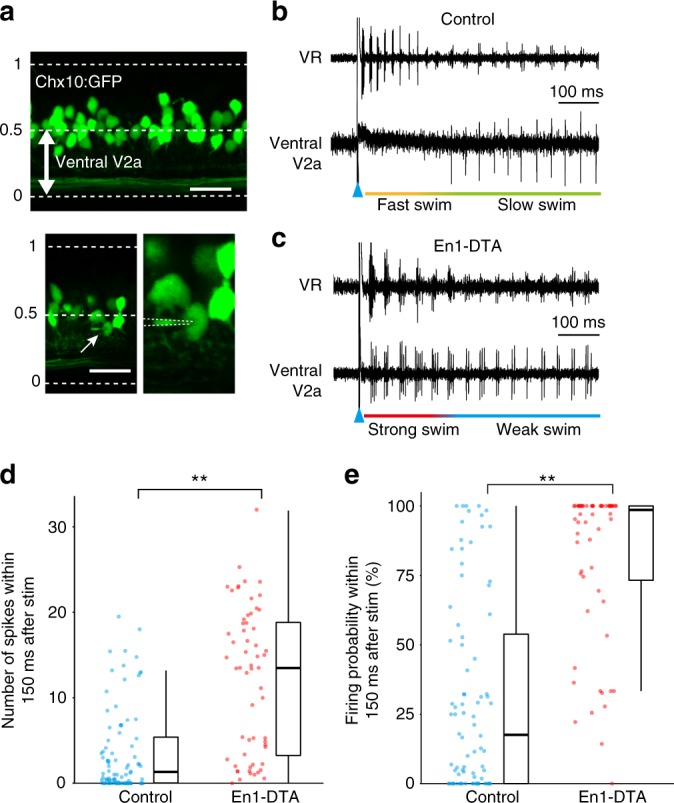
Activity of ventrally located V2a neurons in control and En1-DTA fish. a Lateral view of the spinal cord of Tg[chx10:GFP]. The horizontal dashed lines indicate the dorsal and ventral boundaries of the spinal cord. Ventrally located V2a neurons (the position from 0 to 0.5) are the subjects of the recordings (top panel). The bottom two panels show the images of a loose-patch recording. Scale bar, 20 μm. b An example of simultaneous recordings between ventrally located V2a neurons (loose-patch) and ventral root (VR) in control fish. c An example of simultaneous recordings between ventrally located V2a neurons (loose-patch) and VR in En1-DTA fish. d, e Numbers of spikes (d) and firing probability in each cycle (e) during fast/strong swim. Each circle represents each cell (n = 84 for ventrally located V2a neurons in control; n = 60 for ventrally located V2a neurons in En1-DTA). **P < 0.01 (Mann–Whitney U test, P = 1.3 × 10−10 [d], P = 3.7 × 10−14 [e]). Boxes represent the interquartile range (IQR) between first and third quartiles and the line inside represents the median. Whiskers denote the lowest and highest values within 1.5 × IQR from the first and third quartiles, respectively

Acknowledgments
This image is the copyrighted work of the attributed author or publisher, and ZFIN has permission only to display this image to its users. Additional permissions should be obtained from the applicable author or publisher of the image. Full text @ Nat. Commun.