IMAGE

Figure 5

ID
ZDB-IMAGE-190723-478
Source
Figures for Page et al., 2019
Image
Figure Caption

Figure 5

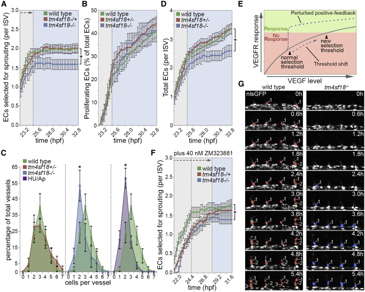
Tm4sf18 Modulates the Magnitude and Timing of the Angiogenic Response

(A and B) Quantification of the number of ECs selected to branch (A) or the percentage of ECs that undergo proliferation (B) in WT, tm4sf18+/−, and tm4sf18−/− embryos (n = 62 ISVs from 16 WT, 58 ISVs from 15 tm4sf18+/−, and 31 ISVs from 8 tm4sf18−/− embryos).

(C) Quantification of the distribution of ISV cellularity in WT, tm4sf18+/−, tm4sf18−/−, and HU/Ap-treated embryos (n = 65 ISVs from 16 WT, 62 ISVs from 15 tm4sf18+/−, 31 ISVs from 8 tm4sf18−/−, and 88 ISVs from 22 HU/Ap-treated embryos).

(D) Quantification of the total number of ECs per ISV in WT, tm4sf18+/−, and tm4sf18−/− embryos. n is the same as in (A).

(E) Predicted shift in the level of VEGF signaling required to achieve a selection threshold in the absence of positive feedback.

(F and G) Quantification of the number of ECs selected to branch in 40 nM ZM323881-treated WT, tm4sf18+/− and tm4sf18−/− embryos (F) and corresponding time-lapse images of sprouting ISVs in 40 nM ZM323881-treated WT and tm4sf18−/− embryos from 20 hpf (G). Embryos were incubated with 40 nM ZM323881 from 18 hpf onward. Nuclei of sprouting ECs emerging from the DA are pseudocolored (n = 26 ISVs from 10 WT, 50 ISVs from 20 tm4sf18+/−, and 21 ISVs from 10 tm4sf18−/− embryos).

Data are means ± SEM. p < 0.05, two-way ANOVA or two-tailed t test. Scale bar, 25 μm.

See also Figure S3.

Acknowledgments
This image is the copyrighted work of the attributed author or publisher, and ZFIN has permission only to display this image to its users. Additional permissions should be obtained from the applicable author or publisher of the image. Full text @ Cell Rep.