IMAGE

Figure 4

ID
ZDB-IMAGE-190723-477
Source
Figures for Page et al., 2019
Image
Figure Caption

Figure 4

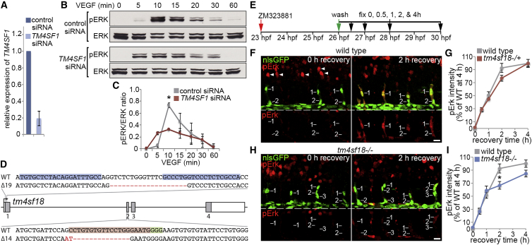
TM4SF1/Tm4sf18 Expression Feeds Back to Amplify VEGF/Vegf Signaling

(A) Relative expression levels of TM4SF1 by qPCR in HUVECs transfected with control or TM4SF1-targeted siRNA (n = 4 separate experiments).

(B and C) Western blot analysis of pERK/ERK levels in HUVECs after VEGF-A stimulation following transfection with control or TM4SF1-targetting siRNA (B) and quantification of pERK/ERK ratios (C) (n = 3 separate experiments).

(D) Lesions introduced into the tm4sf18 loci by TALEN and CRISPR gene editing. A 19-bp deletion of tm4sf18 exon-1 and a 16-bp deletion and 2-bp insertion of exon-2 were generated using TALENs and CRISPR/Cas9, respectively. Genomic target sites for the TALENs, gRNA target site, and PAM sequence are indicated by blue, red, and green highlights, respectively.

(E) Strategy for assessing Vegfr signaling dynamics in vivo.

(F–I) Lateral views of pErk immunostaining in ECs of WT (F) or tm4sf18−/− (H) Tg(kdrl:nlsEGFP)zf109 embryos at 0 and 2 h after inhibitor washout and quantification of pErk fluorescence intensity in WT, tm4sf18+/− (G) or tm4sf18−/− (I) embryos. Arrowheads in (F) indicate pErk in neuronal cells (n = at least 39 ECs from 8 WT, 129 ECs from 20 tm4sf18+/−, and 74 ECs from 13 tm4sf18−/− embryos at each time point).

Data are mean ± SEM. p < 0.05, two-tailed t test. Scale bars, 25 μm.

Acknowledgments
This image is the copyrighted work of the attributed author or publisher, and ZFIN has permission only to display this image to its users. Additional permissions should be obtained from the applicable author or publisher of the image. Full text @ Cell Rep.