IMAGE

Figure 3

ID
ZDB-IMAGE-190723-475
Source
Figures for Page et al., 2019
Image
Figure Caption

Figure 3

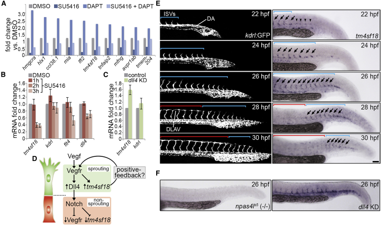
Identification of Putative Positive-Feedback Modulators of Vegf Signaling

(A) Fold change in the indicated transcript levels by microarray following inhibition of Vegfr signaling (2.5 μM SU5416), Notch activity (100 μM DAPT), or both, from 22 to 30 hpf.

(B and C) Fold change in tm4sf18, kdrl, flt4, and dll4 transcript levels by qPCR in embryos incubated with 2.5 μM SU5416 for the indicated times (B) and tm4sf18 and kdrl transcript levels by qPCR upon dll4 KD (C; n = 3 separate experiments).

(D) Illustration of the putative transcriptional regulation of tm4sf18 by Vegf-Notch and proposed function as a positive-feedback modulator of Vegfr signaling.

(E) Lateral views of sprouting ISVs in Tg(kdrl:GFP)s843 embryos (left) or WT embryos following whole-mount in situ hybridization analysis of tm4sf18 expression (right). Blue brackets indicate nascent ISVs; red brackets indicate anastomosed ISVs; arrows indicate tm4sf18-expressing ISVs; and arrowheads indicate tm4sf18 expression at regions of future angiogenic remodeling.

(F) Whole-mount in situ hybridization analysis of tm4sf18 expression in npas4ls5 mutant embryos showing loss of expression, as well as upon dll4 KD showing ectopic expansion of tm4sf18 expression to the DA, consistent with de-repression of Vegfr signaling.

Data are mean ± SEM. Scale bar, 100 μm.

See also Figure S2.

Acknowledgments
This image is the copyrighted work of the attributed author or publisher, and ZFIN has permission only to display this image to its users. Additional permissions should be obtained from the applicable author or publisher of the image. Full text @ Cell Rep.