
Image
|
Figure Caption
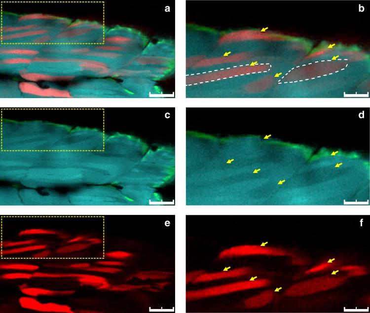
Fig. 3
Single fiber analysis of homology-directed repair (HDR) events. Cross-sectional in vivo imaging of a zebrafish embryo (3 dpf) co-injected with NU7441 at 50 µM and RS-1 at 30 µM.
Acknowledgments
This image is the copyrighted work of the attributed author or publisher, and
ZFIN has permission only to display this image to its users.
Additional permissions should be obtained from the applicable author or publisher of the image.
Full text @ Commun Biol