IMAGE

Figure 7

ID
ZDB-IMAGE-190723-353
Source
Figures for Sharma et al., 2019
Image
Figure Caption

Figure 7

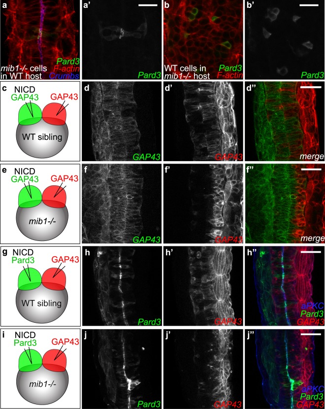
Dissection of the spatial requirement for Notch signaling in neural tube morphogenesis. (a,a’) Pard3-GFP expressing mib1 mutant cells undergo correct polarization when transplanted into WT hosts (n = 30 cells in 6 embryos). (b,b’) In contrast, Pard3-GFP expressing WT cells fail to polarize when transplanted into mib1 mutant hosts (n = 96 cells in 13 embryos). (a,b) are single confocal sections, (a’,b’) maximum projections of 5 slices separated by 2 µm intervals to visualize GFP-positive clones. (c-f) The two halves of the neural tube were labelled by injecting RNAs encoding red or green fluorescent membrane labels (GAP43) into the 2 blastomeres of 2-cell stage embryos (see Methods). (c,d) In WT sibling embryos half-injected with RNA encoding constitutively activated Notch (NICD), cells originating from both sides of the neural tube cross the neural tube midline to integrate the contra-lateral organ half (n = 7/7). (e,f) If NICD is half-injected into mib1 mutants, NICD-containing cells display extensive midline crossing (f,f”), while the crossing of NICD-negative cells is reduced in 6/8 embryos (f’,f”). (g–j) One half of the embryo was injected with RNA encoding NICD Pard3-GFP, the other half with GAP43-RFP. (g,h) 6/6 WT sibling embryos display apical Pard3 accumulation (h) and bilateral midline crossing (h,h’). (i,j) In mib1 mutants, NICD causes apico-basal polarization and midline crossing of Pard3-GFP positive cells in 6/6 embryos (j). NICD-negative GAP43-RFP positive cells fail however to cross the neural tube midline in 5/6 embryos (j’). Pictures represent dorsal views of the spinal cord (anterior up) at 30 somites (a,b), 18 somites (d,f) and 21 somites (h,j) stages. Scalebars: a,b 20 µm, (d,f,h,j) 40 µm.

Acknowledgments
This image is the copyrighted work of the attributed author or publisher, and ZFIN has permission only to display this image to its users. Additional permissions should be obtained from the applicable author or publisher of the image. Full text @ Sci. Rep.