IMAGE

Figure 3

ID
ZDB-IMAGE-190723-348
Source
Figures for Sharma et al., 2019
Image
Figure Caption

Figure 3

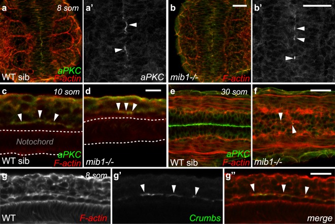
Notch signaling is dispensable for the emergence of floor plate apico-basal polarity. (a,b) At the 8 somites stage, similar discontinuous patches of polarized aPKC are detected in the ventral-most neural tube of WT siblings (a, n = 31) and mib1 mutants (b, n = 14). Dorsal views, anterior up. (a’,b’) are high magnification views of the polarized aPKC signal. (c,d) aPKC is enriched at the apical surface of floor plate cells in 10 somites stage WT siblings (c, n = 8) and mib1 mutants (d, n = 4). Lateral views, anterior left. (e,f) By the 30 somites stage polarized aPKC staining is reduced to few isolated cells in the ventral-most neural tube of mib1 mutants (arrowheads in f, n = 5). Dorsal view, anterior left. (g) Crumbs protein accumulates at the apical surface of 8 somites stage floor plate cells (arrowheads). Lateral view, anterior left. Scalebars: (a,b) 40 µm, (c,d) 10 µm, (eg) 20 µm.

Acknowledgments
This image is the copyrighted work of the attributed author or publisher, and ZFIN has permission only to display this image to its users. Additional permissions should be obtained from the applicable author or publisher of the image. Full text @ Sci. Rep.