IMAGE

FIGURE 3

ID
ZDB-IMAGE-190723-2469
Source
Figures for Antinucci et al., 2018
Image
Figure Caption

FIGURE 3

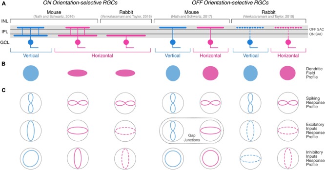
Morphological and physiological features of orientation-selective retinal ganglion cells in mouse and rabbit. Schematic summarizing the morphological (A,B) and physiological (C) properties of ON and OFF OSGCs in mouse (Nath and Schwartz, 2016, 2017) and rabbit (Venkataramani and Taylor, 2010, 2016). Dendritic stratification (A) in the inner plexiform layer (IPL), and planar dendritic field profiles (B) of OSGCs are displayed. Dark gray lines in the IPL indicate ON and OFF choline acetyltransferase (ChAT)-labeled strata corresponding to ON and OFF starburst amacrine cell (SAC) neurites, respectively. The IPL dendritic stratification of rabbit OFF OSGCs is represented with dashed lines because it was not explicitly described by Venkataramani and Taylor (2010), but other studies have reported both mono- and bistratified OFF OSGCs in rabbit (see Table 1). (C) Response profiles of OSGC spiking (top), excitatory inputs (middle), and inhibitory inputs (bottom). Dashed lines of excitatory and inhibitory inputs response profiles in rabbit OSGCs indicate estimated profiles from responses recorded only during preferred and orthogonal orientation stimulation. In mouse OFF OSGCs, the tuned response profiles of gap junction-mediated electrical inputs are indicated (dashed box) instead of excitatory inputs from chemical synapses. INL, inner nuclear layer; GCL, ganglion cell layer. Diagram modified and expanded from Antinucci et al. (2016a) with permission.

Acknowledgments
This image is the copyrighted work of the attributed author or publisher, and ZFIN has permission only to display this image to its users. Additional permissions should be obtained from the applicable author or publisher of the image. Full text @ Front. Neural Circuits