
|
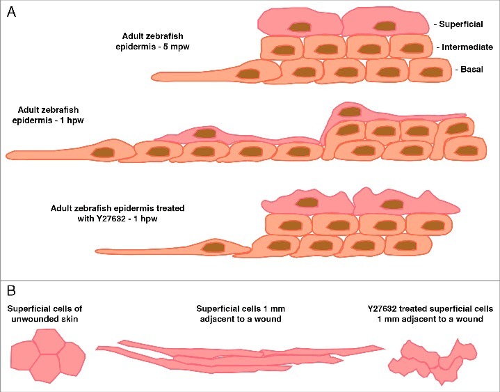
Figure 2.
(A) Representation of a cross section through the epidermis at the wound edge in an adult zebrafish 5 minutes post-wounding (mpw), one hour post-wounding (hpw), or 1 hpw following Rock inhibition. The epidermis initially consists of 2-3 layers of inner basal and intermediary cells and a layer of superficial cells. Within 5 mpw, the LE basal cells have begun to make lamellipodial protrusions and elongate onto the denuded area. By 1 hpw directed radial intercalations of intermediary cells into the basal layer have occurred producing a single layer of inner cells; the superficial cells have elongated to ensure coverage of the increased basal layer area; the LE cells and several cells behind are still protrusive and have undergone some cell elongation, collectively allowing the whole epidermal sheet to move forward to cover the wound. Following Rock inhibition the processes of radial intercalation and coordinated elongation are severely affected resulting in reduced forward movement even though LE protrusions are maintained. (B) Dorsal view outlines of 4 individual superficial cells taken from images from adult zebrafish at the indicated distance from a full-thickness wound (wound is to the left). In unwounded epidermis, the superficial cells are hexagonal in shape. Following wounding the superficial cells elongate in the direction of the wound to up to 10-times their original diameter. Following Rock inhibition the superficial cells change shape but in an uncoordinated manner resulting in many random shapes.