
Image
|
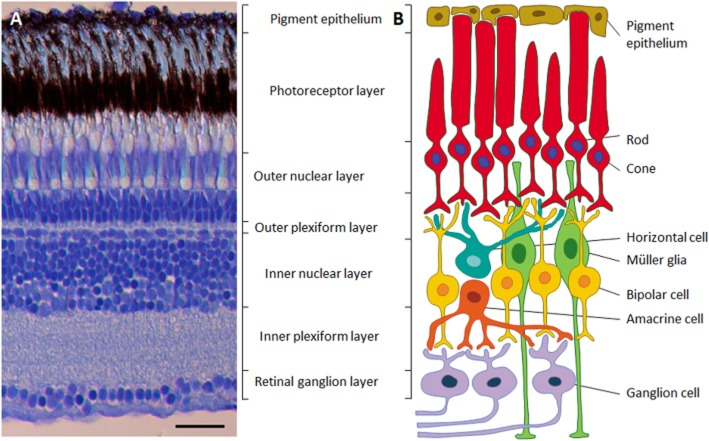
Figure Caption
Figure 1
Structure of the retina. A: Microphotograph of a cross-section through the retina of an adult zebrafish, showing the different cellular and synaptic retinal layers. B: Diagram of the neural circuit of the retina, showing the six neuronal cell types and the two supporting cell types (Müller glia and retinal pigmented epithelium). In A, the scale bar = 25μm.
Acknowledgments
This image is the copyrighted work of the attributed author or publisher, and
ZFIN has permission only to display this image to its users.
Additional permissions should be obtained from the applicable author or publisher of the image.
Full text @ Br. J. Pharmacol.