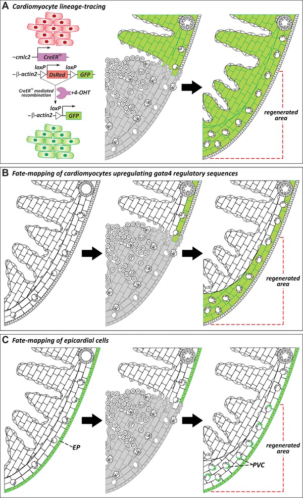
IMAGE

Figure 3

ID
ZDB-IMAGE-190723-2213
Source
Figures for González-Rosa et al., 2017
Image
Figure Caption

Figure 3

Cellular origins of regenerated tissue. (A) Genetic lineage‐tracing experiments to determine the origin of regenerated myocardium during zebrafish heart regeneration. Virtually all cardiomyocytes in uninjured hearts are labeled by GFP expression after treatment with 4‐hydroxytamoxifen (4‐OHT). In regenerated hearts, the new myocardium is GFP+, revealing that new muscle derives from the proliferation of preexisting cardiomyocytes (Jopling et al., 2010; Kikuchi et al., 2010). (B) A population of subepicardial cardiomyocytes activates regulatory sequences of gata4 upon injury. Fate mapping of cardiomyocytes that activate gata4 regulatory sequences reveals that these cells contribute preferentially to myocardial regeneration (Kikuchi et al., 2010). (C) Lineage tracing of epicardial cells using tcf21:CreERT2 (Kikuchi, Gupta, et al. 2011) or tissue transplants (González‐Rosa et al., 2012) demonstrates that the epicardium gives rise to perivascular cells but not myocardium in the regenerated heart. EP, epicardium; PVC, perivascular cell

Acknowledgments
This image is the copyrighted work of the attributed author or publisher, and ZFIN has permission only to display this image to its users. Additional permissions should be obtained from the applicable author or publisher of the image. Full text @ Regeneration (Oxf)