Figure Caption
Figure 1
A brief summary of zebrafish experimental models in neuroscience researchPanel A shows the evolving nature of zebrafish models in the last 50 years, initially used mainly for basic genetic and neurodevelopmental studies, but more recently applied to developing in-vivo models for complex brain disorders, such as autism, depression and psychoses. Inset: selected zebrafish strains useful in biological psychiatry research (top to bottom: adult wild type zebrafish, casper, spiegeldanio and nacre mutants); photos courtesy of the Kalueff (ZENEREI Institute, USA), the Norton (University of Leicester, UK), the Parichy (University of Washington, USA) laboratories and Carolina Biological Supply Company (Burlington, USA).
Panel B illustrates two research strategies which can both be applied to zebrafish models. As a vertebrate species amenable to in-vivo analyses and with high genetic/physiological homology to humans, zebrafish are ideal for ‘intensive’, mechanistically driven neuroscience research into conserved, core molecular pathways or neural circuits (photo). Due to their small size, ease of maintenance and short generation time, zebrafish also represent an excellent model for ‘extensive’ biomedical research, including low-cost high-throughput screening for small molecules or genetic mutations. Both strategies can lead to the development of novel therapies for major psychiatric disorders. Photo: visualizing zebrafish signaling as an example of intensive, pathway-oriented mechanistic research (the left image shows expression patterns of genetically encoded calcium indicator; zebrafish at 5 dpf show pan-neuronal expression of HuC:Gal4; UAS:GCaMP5, a zoomed-in image on the right shows a single cell resolution: note that some cells are activated, as indicated by the increased fluorescence; scale bar 100 µm).
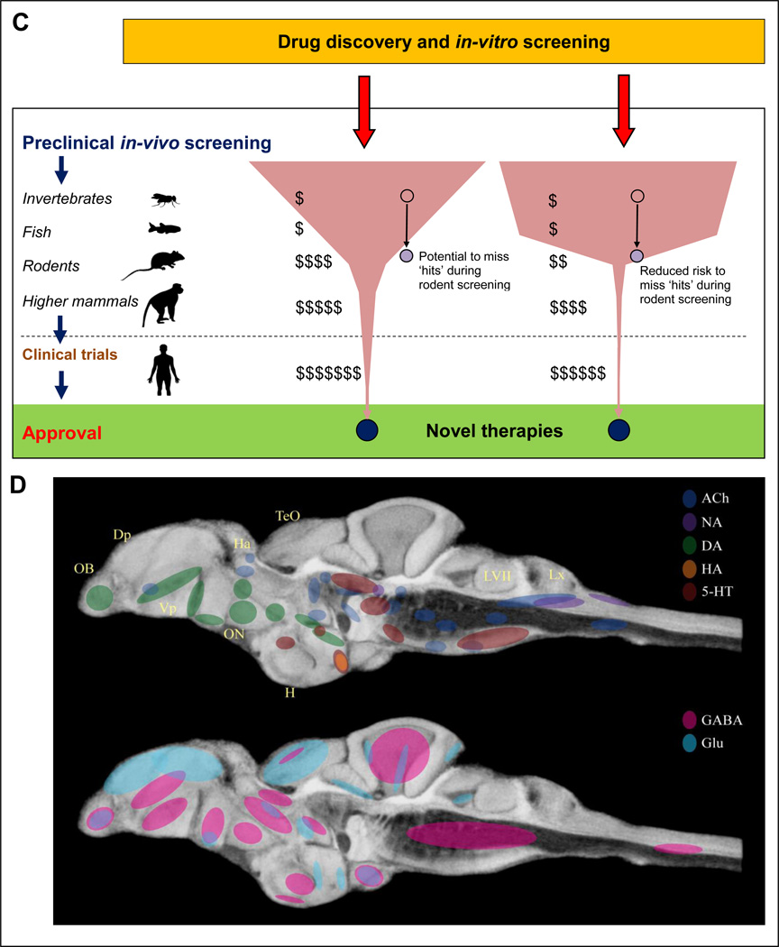
Panel C illustrates the benefits of including inexpensive zebrafish models into preclinical screening batteries. A substantial fiscal saving (shown as $) can be achieved by narrowing screening to potentially active compounds using fish (rather than mice) as the first vertebrate model organism in the screening pipeline. In addition, the risks of missing promising ‘hits’ are lower because the subsequent screening in mammals will be ‘confirmatory’, and based on more valid data generated from zebrafish (rather than invertebrate or in-vitro) tests.
Panel D summarizes major neurotransmitter systems in the adult zebrafish brain: ACh - acetylcholine (dark blue), NA - noradrenergic (purple), DA - dopamine (green), HA - histamine (orange), 5-HT - serotonin (red); GABA - γ-aminobutyric acid (pink), Glu - glutamate (light blue). The highlighted brain regions include olfactory bulb (OB), dorsal pallium (Dp), ventral pallium (Vp), habenula (Ha), optic nerve (ON), facial lobe (LVII) and vagal lobe (LX). Highlighted areas have been overlaid over a minimum deformation model of the adult zebrafish brain.
Acknowledgments
This image is the copyrighted work of the attributed author or publisher, and
ZFIN has permission only to display this image to its users.
Additional permissions should be obtained from the applicable author or publisher of the image.
Full text @ Mol. Psychiatry