
|
Figure 1
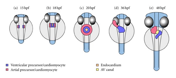
Stages of heart development in zebrafish embryos in a ventral view. (a) At 15 hpf cardiac precursors move towards the anterior lateral plate mesoderm; the atrial precursors are located laterally, the ventricular precursors medially. (b) Cardiac fusion starts at about 18 hpf at the posterior end of the bilateral heart fields; first, endocardial cells arrive at midline, followed by ventricular cells. The atrial precursors do so slightly later. (c) After cardiac fusion the cells form the cardiac cone. Viewed ventrally, this structure resembles a ring: endocardial cells lining the central lumen, and ventricular cells are surrounded by atrial precursors. The cardiac cone starts to transform into the heart tube by 21 hpf. (d) Cardiac looping of the heart tube occurs between 26 hpf and 48 hpf. The linear heart tube bends and creates an S-shaped loop. By 36 hpf the atrium and ventricle become distinguishable. (e) The heart tube rotates. The different parts of the heart tube do not rotate equally and therefore torsion occurs. The AV canal develops by constriction of the boundary between atrium and ventricle. Ventricular maturation by appositional growth takes place.