IMAGE

Figure 2

ID
ZDB-IMAGE-190723-1913
Source
Figures for Dimitriadi et al., 2018
Image
Figure Caption

Figure 2

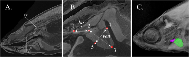
Multiple views from a single scan of an adult zebrafish. (A) Representative oblique slice defined by three landmarks (anterior and posterior end of the bulbus arteriosus, the centre of the 1st vertebra). (B) Inset of Fig. 2A, showing the ventricle (ven), bulbus arteriosus (ba), as well as the distance measurements taken. (C) Three-dimensional volume rendering of the bulbus arteriosus (purple) and ventricle (green). V, first vertebra. ba, bulbus arteriosus. ven, ventricle. Landmark 1, bulbus junction with the first branchial arch. Landmark 2, ventricle – bulbus valve. Landmark 3, ventricle apex. Landmarks 4 and 5 define the widest distance of the ventricle, perpendicularly to ventricle length (D2–3). D1–2, Bulbus-arteriosus length (BaL); D2–3, Ventricle length (VL); D4–5, maximum ventricle depth (VD), perpendicular to VL.

Acknowledgments
This image is the copyrighted work of the attributed author or publisher, and ZFIN has permission only to display this image to its users. Additional permissions should be obtained from the applicable author or publisher of the image. Full text @ Sci. Rep.